SpudCell FAQ

What is SpudCell

SpudCell is a liposome encapsulated synthetic cell that can grow and replicate.

It undergoes a full cell cycle, with DNA replication, genetically encoded feeding and genetically encoded division. 

SpudCell manual

Protocols for SpudCell formation and experiments

Below is the list of protocols and procedures needed to assemble and analyze a SpudCell.


Read this first

Synthetic cell experiments are still more resource-intensive than growing natural cells. While our motivation for this research is to make biology a general purpose technology, usable freely by all, we are currently operating in the sandbox environment.

To grow a SpudCell, your lab needs to have core competencies in:

  • liposome preparation
  • PURE system expression
  • aHL expression in liposomes

If you haven’t used any of those techniques before, we strongly suggest you successfully set up those first, before moving on to SpudCell experiments.


pREP Transcription–Translation and Phi29 Replication Reaction

  1. Purpose

This protocol describes the preparation and incubation of the pREP cell-free transcription–translation and DNA replication system used for protein expression, Phi29 genome replication, and related synthetic cell experiments.

  1. Materials

2.1 DNA preparation

  • Plasmid DNA (mini-prepped, Epoch 2160250)
  • PCR clean-up kit (Epoch 2360250)
  • Lyophilizer for DNA concentration

2.2 Commercial reagents

  • PURExpress Solution A (NEB E6800L)
  • PURExpress Solution B (NEB E6800L)
  • NxGen Phi29 DNA polymerase (Lucigen 30221-2)
  • DTT (Goldbio DTT50)
  • dNTPs (Denville Scientific CB4420-2)
  • Amino acids (Sigma-Aldrich)
  • Creatine phosphate (Sigma-Aldrich 27920-5G)
  • Spermidine (Sigma-Aldrich S2526-5G)
  • tRNA mix (E. coli, Sigma-Aldrich 10109550001)
  1. Stock Solutions (store at −80 °C)

3.1 10× Energy Mix

Prepare and aliquot:

  • 700 mM potassium glutamate
  • 79 mM magnesium glutamate
  • 1 M HEPES pH 8.0
  • 250 mM creatine phosphate
  • 3.75 mM spermidine
  • 5.18 g/L E. coli tRNAs
  • 60 mM DTT
  • 3.6 mM each L-amino acid (20 standard amino acids)

3.2 50× rNTP Mix

Prepare and aliquot:

  • 18.75 mM ATP
  • 12.5 mM GTP
  • 6.25 mM CTP
  • 6.25 mM UTP
  1. Reaction Setup

4.1 Final reaction composition (per 100 µL reaction)

Prepare reactions on ice.

Component

Final concentration

Volume (per 100 µL)

10× Energy Mix

10 µL

50× rNTP Mix

2 µL

PURExpress Solution A

0.1×

2 µL

PURExpress Solution B

60% v/v

60 µL

dATP

0.6 mM

variable (from stock)

dGTP

0.6 mM

variable

dCTP

0.6 mM

variable

dTTP

0.6 mM

variable

DTT

4 mM

variable

Phi29 DNA polymerase

0.4 U/µL

variable

Plasmid DNA

as required

variable

Nuclease-free water

to 100 µL

  1. Master Mix Preparation (recommended workflow)

5.1 Prepare bulk reaction master mix (excluding DNA and Phi29 polymerase if variable)

For N reactions, prepare:

  1. Combine in a sterile tube on ice:
    • 10× Energy Mix (1× final)
    • 50× rNTP Mix (1× final)
    • PURExpress Solution A (0.1× final)
    • PURExpress Solution B (60% final)
    • dNTPs (0.6 mM each final)
    • DTT (4 mM final)
    • Nuclease-free water (to ~90% of final volume)
  2. Mix gently by pipetting (do not vortex).
  3. Keep on ice until use.

5.2 Final assembly per reaction

For each 100 µL reaction:

  1. Aliquot 90–95 µL of master mix into PCR tubes or plate wells.
  2. Add:
    • Plasmid DNA (variable, typically 10–500 ng depending on experiment)
    • Phi29 DNA polymerase to 0.4 U/µL final concentration
  3. Mix gently by pipetting.
  4. Briefly spin down (5–10 s).
  1. Incubation
  1. Incubate reactions at 30 °C.
  2. Standard reaction time: 8 hours.
  3. Avoid agitation unless specifically required for experimental variation.
  1. Storage and handling notes
  • Keep all reagents on ice during setup.
  • Avoid repeated freeze–thaw cycles of energy and nucleotide mixes.
  • Prepare aliquots of master mixes when running multiple reactions.
  • DNA should be highly purified and concentrated prior to use.


Plasmid Preparation, Cloning, and Usage for pREP / In Vitro Translation Experiments

  1. Purpose

This protocol describes plasmid sourcing, cloning strategies, purification workflow, and concentration handling for DNA templates used in pREP transcription–translation and related in vitro expression systems.

  1. Plasmid Sources

2.1 External plasmid sources (non-lab-cloned constructs)

  • pLD1, pLD2, pLD3 plasmids (generous gift from Dr. Anthony Forster)
  • α-hemolysin (αHL) and T7 RNA polymerase plasmids (generous gift from Dr. Vincent Noireaux)

2.2 Promoter system standardization

  • All plasmids under T7Max promoter were cloned into a pre-existing lab vector system (previously described in ref. 67).
  • This backbone contains:
    • UTR1 sequence (optimized for bacterial in vitro translation)
    • T500 transcription terminator (optimized for bacterial in vitro translation)
  1. Cloning Procedures

3.1 General cloning methods used
All cloning was performed using one of the following:

  • Gibson Assembly
    • NEB Gibson Assembly Master Mix (E2611)
  • Site-directed mutagenesis
    • Q5 High-Fidelity DNA Polymerase (NEB M0493)
    • KLD Enzyme Mix (NEB M0554)
  1. Plasmid Amplification

4.1 Bacterial host strain

  • DH5α E. coli cells used for plasmid amplification

4.2 Workflow

  1. Transform plasmid constructs into DH5α cells
  2. Grow under standard antibiotic selection conditions
  3. Isolate plasmid DNA from overnight cultures
  1. Plasmid Purification (Double-Clean Workflow)

5.1 Primary purification

  • GenCatch™ Plasmid DNA Mini-Prep Kit (Epoch 2160250)

5.2 Secondary purification

  • GenCatch™ PCR Cleanup Kit (Epoch 2360250)

5.3 Rationale

  • Double purification was used based on empirical observations (colleagues’ reports) indicating improved in vitro translation yield with highly purified DNA templates.
  1. DNA Quantification and Normalization

6.1 Standard reaction concentrations

  • Typical plasmid concentration in translation reactions:
    • 10 nM final concentration per plasmid

6.2 Multi-plasmid conditions

  • When multiple plasmids were used:
    • Each plasmid was added at equal molar concentration
    • Exception:
      • Whole 90 kb genome experiments used 2.5 nM per plasmid
  1. DNA Handling for Multi-Plasmid Reactions

7.1 Problem addressed

  • pREP reaction volume constraints result in limited free water volume after reagent addition.

7.2 Solution for high-plasmid load (>3 plasmids)

  1. Combine plasmid stock solutions into a single tube
  2. Lyophilize combined DNA mixture
  3. Resuspend concentrated DNA in minimal volume of nuclease-free water
    • Typical resuspension volume: ~3 µL for a 30 µL final reaction

7.3 Outcome

  • Enables high-complexity DNA inputs without increasing total reaction volume or diluting other components
  1. Standard Use in pREP Reactions

8.1 DNA input integration

  • Add plasmid DNA to final reaction mix at desired molarity (typically 10 nM per construct unless specified otherwise)

8.2 Mixing strategy

  • For multi-plasmid experiments:
    • Pre-mix DNA stocks prior to reaction setup when necessary
    • Ensure equal molar representation unless experimental design specifies otherwise
  1. Storage and Handling Notes
  • Store plasmid stocks at −20 °C
  • Avoid repeated freeze–thaw cycles
  • Use double-purified DNA for optimal in vitro expression performance
  • Maintain concentrated DNA stocks for multi-plasmid systems to minimize dilution effects


Synthetic Cell Liposome Preparation (Oil Extrusion Method)

  1. Purpose

This protocol describes the preparation, purification, and handling of synthetic cell liposomes used in cell-free synthetic biology experiments. Liposomes are prepared using a lipid thin-film method followed by oil hydration, emulsification, and differential centrifugation purification.

  1. Materials

2.1 Lipids

  • DOPE (1,2-dioleoyl-sn-glycero-3-phosphoethanolamine; Avanti 850725P)
  • DOPC (1,2-dioleoyl-sn-glycero-3-phosphocholine; Avanti 850375P)
  • Cholesterol (Avanti 700100P)

2.2 Solutions

  • Chloroform (anhydrous, for lipid stocks)
  • Mineral oil
  • Centrifuge buffer:
    • 100 mM HEPES
    • 200 mM glucose
    • pH 8.0
  • Wash buffer:
    • 100 mM HEPES
    • 250 mM glucose
    • pH 8.0
  • External reaction buffer (as required per experiment)

2.3 Equipment

  • Glass vials (amber preferred)
  • Glass syringes (for chloroform handling)
  • Nitrogen gas supply
  • Bath sonicator
  • Vortexer with continuous shaking attachment
  • Refrigerated centrifuge (4 °C capable)
  • Gel loading pipette tips (preferred for oil removal)
  1. Lipid Composition

3.1 Standard formulation (“1× liposomes”)

  • Total lipid concentration: 1 mM (excluding cholesterol from total lipid calculation)
  • Molar ratio (DOPE:DOPC:Cholesterol): 1:3:1

3.2 Final composition per 1 mM lipid sample

  • DOPE: 0.25 mM
  • DOPC: 0.75 mM
  • Cholesterol: 0.25 mM
  1. Preparation of Lipid Thin Film

4.1 Preparation of stock solutions

  • Prepare lipid stocks in chloroform at 1 mg/mL

4.2 Thin film formation (typical 30 µL liposome batch)

  1. In an amber glass vial, combine:
    • 5.58 µL DOPE stock
    • 17.7 µL DOPC stock
    • 2.9 µL cholesterol stock
  2. Mix using glass syringes only (avoid plastic contact with chloroform).
  3. Evaporate chloroform under gentle nitrogen stream for 6 hours to form a dry lipid film.
  4. Cap vials tightly and store at −20 °C until use.
  1. Liposome Formation (Oil Hydration and Emulsification)

5.1 Lipid hydration in oil phase

  1. Add dried lipid film to 200 µL mineral oil
  2. Incubate at 60 °C for 3 hours
    • Vigorously shake during incubation
  3. Sonicate in bath sonicator for 10 minutes
  4. Incubate overnight at room temperature
    • Continuous vigorous shaking (vortexer with attachment)
  5. Cool mineral oil suspension to 4 °C
    • Maintain shaking during cooling
  1. Liposome Emulsification and Encapsulation

6.1 Emulsion formation

  1. Add 30 µL internal aqueous solution to mineral oil lipid suspension
  2. Vortex vigorously for 5 minutes at 4 °C
  1. Gradient Purification by Centrifugation

7.1 First centrifugation (phase separation)

  1. Carefully layer emulsion onto:
    • 250 µL centrifuge buffer (100 mM HEPES, 200 mM glucose, pH 8)
  2. Centrifuge at:
    • 18,000 × g (rcf), 4 °C, 15 minutes
  3. After spin:
    • Carefully remove oil phase using gel loading tip

7.2 Liposome recovery and wash

  1. Collect liposomes from bottom aqueous phase using fresh pipette tip
  2. Transfer to:
    • 250 µL pre-chilled wash buffer (100 mM HEPES, 250 mM glucose, pH 8)
  3. Centrifuge:
    • 12,000 × g (rcf), 4 °C, 5 minutes

7.3 Final cleanup

  1. Remove any residual oil layer from top of wash buffer
  2. Transfer liposome pellet/phase to fresh tube
  3. Resuspend in final external reaction buffer
  4. Store or proceed immediately to experiments
  1. Temperature Control
  • All steps from emulsification onward must be performed at 4 °C
  • Maintain samples in cold room or on ice as appropriate
  1. Liposome Size Control (Extrusion Parameters)

Liposome size is defined by extrusion membrane pore size during initial lipid processing:

  • 2 µm membrane → synthetic cell cycle experiments
  • 0.8 µm membrane → DLS characterization samples
  • 0.4 µm membrane → feeder liposomes
  1. Storage and Handling Notes
  • Lipid stocks stored in chloroform must be kept in amber vials
  • Thin lipid films stored at −20 °C
  • Avoid repeated freeze–thaw of lipid films
  • Use gel-loading tips for oil removal to maximize phase precision
  • Prevent lipid oxidation by minimizing light exposure

Fluorescent Labeling of Synthetic Cell Liposomes

  1. Purpose

This protocol describes incorporation of fluorescent membrane dyes into lipid thin films for preparation of labeled synthetic cell liposomes used in imaging and tracking experiments.

  1. Materials

2.1 Lipid dyes (chloroform stocks)

All dyes are used as 100 µg/mL stocks in chloroform:

  • NBD-PE
    • (N-(7-Nitrobenz-2-Oxa-1,3-Diazol-4-yl)-1,2-dihexadecanoyl-sn-glycero-3-phosphoethanolamine; Thermo Fisher N360)
  • Rhodamine-PE (Lissamine™ Rhodamine B PE)
    • (1,2-dihexadecanoyl-sn-glycero-3-phosphoethanolamine; Thermo Fisher L1392)
  • Cy5-PE
    • (1,2-dioleoyl-sn-glycero-3-phosphoethanolamine-N-(Cyanine 5); Avanti 810335C)
  • Cy7-PE
    • (1,2-dioleoyl-sn-glycero-3-phosphoethanolamine-N-(Cyanine 7); Avanti 810337C)
  1. Labeling Strategy

3.1 Standard dye incorporation

  • Dye concentration in liposomes:
    • 0.1 mol% per dye relative to total lipid (excluding cholesterol from total lipid calculation)

3.2 Total dye loading rules

  • Single dye system:
    • 0.1 mol% total dye
  • Dual dye system:
    • 0.1 mol% + 0.1 mol% = 0.2 mol% total dye

3.3 Effective concentration example

  • For 1 mM liposomes:
    • 0.1 mol% dye = 1 µM dye per lipid population
  1. Incorporation into Lipid Thin Film

4.1 General procedure

  1. During lipid thin film preparation (see liposome SOP section 4), add dye stock directly into chloroform lipid mixture.
  2. Mix dye stock with lipid chloroform solutions before solvent evaporation step.
  3. Proceed with nitrogen drying and thin film formation as standard.
  1. Dye Stock Addition (30 µL, 1 mM liposome batch)

For a standard 30 µL, 1 mM lipid preparation, add the following dye stock volumes directly into the chloroform lipid mixture:

Dye

Stock concentration

Volume per thin film

NBD-PE

100 µg/mL (chloroform)

2.86 µL

Rhodamine-PE

100 µg/mL (chloroform)

4.0 µL

Cy5-PE

100 µg/mL (chloroform)

3.79 µL

Cy7-PE

100 µg/mL (chloroform)

3.98 µL

  1. Thin Film Formation with Dye
  1. Combine lipid chloroform stocks as per standard liposome preparation protocol.
  2. Add appropriate dye stock volume (from Section 5) directly into lipid chloroform mixture.
  3. Mix thoroughly using glass syringes.
  4. Proceed with:
    • Nitrogen drying (6 hours, gentle flow)
    • Vacuum-free storage at −20 °C until use (sealed amber vial)
  1. Liposome Formation (Downstream Compatibility)

After dye incorporation, liposomes are prepared using the standard oil extrusion protocol:

  • Dye-labeled lipid films are hydrated in mineral oil
  • Follow identical emulsification, centrifugation, and purification steps as non-labeled liposomes
  1. Storage and Handling Notes
  • Dye–lipid mixtures in chloroform must be protected from light (use amber vials)
  • Avoid repeated freeze–thaw of lipid films
  • Minimize oxygen exposure during drying to prevent dye degradation
  • Store dried thin films at −20 °C in dark conditions
  • All dye handling should be performed using glass syringes where possible

Preparation of Feeder Liposomes for Synthetic Cell Growth and Division Experiments

  1. Purpose

This protocol describes the preparation of Ni-NTA–functionalized feeder liposomes used in synthetic cell growth, division, and generation-tracking experiments. Feeder liposomes are designed to deliver biochemical components and enable membrane-mediated interactions such as fusion and cargo exchange.

  1. Materials

2.1 Lipids

  • DOPE (Avanti 850725P)
  • DOPC (Avanti 850375P)
  • Cholesterol (Avanti 700100P)
  • Ni-NTA lipid:
    • 1,2-dioleoyl-sn-glycero-3-[(N-(5-amino-1-carboxypentyl)iminodiacetic acid)succinyl] nickel salt
    • Avanti 790404P

2.2 Lipid stock solutions

  • Lipids prepared in chloroform (standard stocks)
  • Ni-NTA lipid stock:
    • 0.1 mg/mL in chloroform

2.3 Equipment

  • Avanti mini-extruder (Avanti 610023)
  • Polycarbonate membranes (0.4 µm; Avanti 610007)
  • Orbital shaker (4 °C compatible)
  • Cold room (4 °C)
  • Standard liposome preparation setup (see liposome SOP section 4)
  1. Lipid Composition

3.1 Base composition (all feeder liposomes)

  • DOPE:DOPC:Cholesterol = 1:3:1 molar ratio
  • Cholesterol is excluded from total lipid calculations

3.2 Functional lipid addition

  • Ni-NTA lipid added at experiment-specific mol% relative to total lipid (excluding cholesterol)

3.3 Example calculation

  • For 2 mM total lipid liposomes with 0.6 mol% Ni-NTA:
    • Ni-NTA concentration = 12 µM
  1. Thin Film Preparation

4.1 Lipid mixture preparation

  1. Prepare lipid chloroform mixtures as per standard liposome thin film protocol.
  2. Add Ni-NTA lipid stock directly into chloroform lipid mixture prior to drying.

4.2 Example (30 µL, 2 mM feeder liposomes, 0.6 mol% Ni-NTA)

  • Add 3.8 µL Ni-NTA chloroform stock (0.1 mg/mL) to lipid mixture.

4.3 Thin film formation

  1. Mix all lipid components in chloroform using glass syringes.
  2. Dry under gentle nitrogen flow to form thin film.
  3. Store dried film at −20 °C until use.
  1. Liposome Formation

5.1 Standard formation

  • Prepare liposomes using the standard oil extrusion method (see liposome SOP section 4).

5.2 Hydration and emulsification

  • Hydrate lipid film in mineral oil
  • Proceed with heating, sonication, and shaking steps as in standard protocol
  1. Liposome Extrusion

6.1 Purpose

  • Define feeder liposome size for controlled fusion and uptake behavior

6.2 Procedure

  1. Load liposome suspension into Avanti mini-extruder
  2. Extrude through 0.4 µm polycarbonate membranes
  3. Perform 9 extrusion passes
  4. Collect extruded liposomes under sterile cold conditions
  1. Post-Extrusion Equilibration
  1. Transfer liposomes to fresh tube
  2. Incubate at 4 °C for 10 minutes
  3. Gently shake on orbital shaker during equilibration
  4. Proceed immediately to experimental use
  1. Functional Loading for Cell Cycle Experiments

Each feeder liposome population may include:

  • Ni-NTA lipid: 0.6 mol% (typical value unless otherwise stated)
  • Generation counter oligo:
    • 4 nM of one “bottom” oligonucleotide (see Methods section 12)
  • Full pREP reaction mixture:
    • Complete 1× pREP system (see Methods section 2)
  1. Standard Usage Conditions
  • Typical reaction volume per condition:
    • 30 µL feeder liposomes
  • Lipid concentration:
    • 2 mM total lipid
  • Used per generation in growth/division experiments unless otherwise specified
  1. Temperature and Handling
  • All preparation and post-processing steps performed at 4 °C
  • Maintain samples in cold room when possible
  • Avoid prolonged warming after extrusion to preserve functionality
  1. Storage and Stability Notes
  • Dried lipid films stored at −20 °C
  • Ni-NTA lipid is sensitive to repeated freeze–thaw cycles
  • Extruded liposomes should be used shortly after preparation for optimal performance
  • Avoid extended storage after loading with biochemical components

Liposome Fusion Experiments 

  1. Purpose

This protocol describes the preparation, tagging, mixing, incubation, and analysis of synthetic cell liposome fusion experiments using functionalized liposomes (e.g., protein tags, Ni-NTA systems, and αHL-mediated fusion).

  1. Materials

2.1 Liposomes

  • Standard synthetic cell liposomes prepared via:
    • Oil extrusion method (see liposome SOP section 4)
    • Feeder liposome protocol (see feeder SOP section 6)
  • Optional functionalized liposomes:
    • Ni-NTA–functionalized liposomes
    • Fluorescently labeled liposomes

2.2 Fusion tags and reagents

  • Fusion tag systems (prepared according to published protocols75)
  • α-hemolysin (αHL) system (expression-based fusion tag)
  • Vamp-2 fusion proteins (see dedicated section in Methods)

2.3 Chemicals

  • Triton X-100 (Acros Organics AC215682500)

2.4 Equipment

  • Orbital shaker (room temperature and 30 °C capable incubator setup)
  • PCR tubes
  • 96-well plates (for endpoint fluorescence readout)
  • Fluorescence plate reader
  • Incubator set to 30 °C
  1. Liposome Preparation (Pre-Fusion)

3.1 Base preparation

  • Liposomes prepared as described in:
    • Standard liposome SOP (Methods section 4)
    • Feeder liposome SOP (Methods section 6)

3.2 Standard concentration

  • All fusion samples prepared at:
    • 1 mM total lipid concentration
  • Exceptions:
    • Only as specified in experimental variations
  1. Fusion Tag Functionalization

4.1 External fusion tag decoration

  1. Prepare liposomes as described above.
  2. Incubate liposomes with fusion tags (per protocol75).
  3. Incubation conditions:
    • 30 minutes at room temperature
    • Gentle tumbling on orbital shaker

4.2 αHL-based fusion system

  1. Prepare synthetic cells expressing αHL as described in expression protocols.
  2. Incubation conditions:
    • 3 hours at 30 °C
    • Gentle shaking on orbital shaker inside incubator
  1. Sample Mixing for Fusion Assays

5.1 Standard mixing procedure

  1. After tag incubation or αHL expression:
    • Mix liposome populations in equal volumes, unless otherwise specified
  2. Each liposome population is typically:
    • 1 mM total lipid

5.2 Mixing format

  • Perform mixing in:
    • PCR tubes
  • Use gentle pipetting to avoid mechanical rupture
  1. Fusion Incubation

6.1 Standard conditions

  • Incubate mixed samples:
    • 30 °C
    • Gentle shaking on orbital shaker inside incubator
  • Standard incubation time:
    • 6–12 hours (depending on experiment)
      • Typical default: 12 hours
  1. Post-Fusion Analysis

7.1 Fluorescence measurement

  1. Transfer samples to 96-well plates
  2. Measure fluorescence using plate reader

7.2 Molecular analysis (if required)

  • For RT-PCR, qPCR, or Western blot:
    1. Lyse liposomes using:
      • 0.1% Triton X-100
    2. Proceed with downstream molecular assays
  1. Standard Reaction Conditions Summary
  • Lipid concentration:
    • 1 mM total lipid per sample
    • Includes Ni-NTA and/or dyes when applicable
  • Mixing ratio:
    • Typically 1:1 volume ratio
  • Incubation temperature:
    • 30 °C
  • Shaking:
    • Gentle orbital shaking throughout incubation
  1. Storage and Handling Notes
  • Prepare fusion samples fresh when possible
  • Maintain gentle handling to prevent premature rupture
  • Avoid vigorous vortexing after fusion tag binding
  • Keep samples at 30 °C only during incubation phases

FRET-Based Membrane Growth and Lipid Mixing Assay

  1. Purpose

This protocol describes fluorescence resonance energy transfer (FRET)–based quantification of membrane growth and lipid mixing in synthetic cell liposomes. The assay monitors dilution of membrane dyes upon liposome fusion and membrane expansion.

  1. Principle

Membrane growth is quantified by measuring dilution of FRET pairs embedded in lipid bilayers. Upon fusion with unlabeled membranes, FRET efficiency decreases, leading to increased donor fluorescence.

  • Dye dilution → decreased FRET → increased donor signal
  • Signal is calibrated to % lipid mixing using defined standards
  1. Materials

3.1 Lipid dyes (FRET pairs)

  • NBD–Rhodamine pair:
    • NBD-PE (donor)
    • Lissamine Rhodamine-PE (acceptor)
  • Cy5–Cy7 pair:
    • Cy5-PE (donor)
    • Cy7-PE (acceptor)

3.2 Liposomes

  • Prepared via standard liposome thin film and extrusion methods:
    • Liposome SOP (section 4)
    • Fluorescent labeling SOP (section 5)

3.3 Equipment

  • Avanti mini-extruder (Avanti 610023)
  • 0.4 µm polycarbonate membranes (Avanti 610007)
  • Fluorescence plate reader or spectrofluorometer
  • Standard incubation equipment
  1. FRET Pair Selection Rules

4.1 Pair usage

  • NBD–Rhodamine
    • Used when no GFP signal is present
  • Cy5–Cy7
    • Used when GFP is present (avoids spectral overlap with NBD)
  1. Experimental Design

5.1 Liposome populations

  • Population A:
    • Labeled with both donor and acceptor FRET dyes
  • Population B:
    • Unlabeled (no membrane dyes)

5.2 Mixing principle

  • Fusion or membrane growth leads to:
    • Dilution of FRET dyes
    • Decreased FRET efficiency
    • Increased donor fluorescence
  1. FRET Liposome Preparation
  1. Prepare lipid thin films (see liposome SOP section 4)
  2. Incorporate FRET dyes during chloroform stage (see dye labeling SOP section 5)
  3. Form liposomes using standard oil extrusion method
  4. Extrude through:
    • 0.4 µm polycarbonate membranes
    • Using Avanti mini-extruder
    • Perform standard extrusion procedure
  1. FRET Calibration Standards

7.1 Purpose

  • Convert fluorescence signal into % lipid mixing

7.2 Calibration series
Prepare control liposomes at defined dye dilution levels:

  • 0% membrane mixing
    • 1 mM liposomes
    • 0.1 mol% donor + 0.1 mol% acceptor (total 0.2 mol% FRET dyes)
  • 50% membrane mixing
    • 1 mM liposomes
    • 0.05 mol% donor + 0.05 mol% acceptor
  • Additional calibration points prepared analogously by proportional dilution of both dyes

7.3 Preparation method

  • All calibration samples:
    • Prepared from thin lipid films
    • Extruded through 0.4 µm membranes (as above)
  1. FRET Measurement

8.1 Signal acquisition

  • Measure fluorescence using donor-specific wavelengths:
    • NBD or Cy5 emission channels depending on system used

8.2 Spectral configuration

  • Refer to Table S5 for excitation/emission settings
  1. Data Interpretation

9.1 Signal readout

  • Increased donor fluorescence indicates:
    • Reduced FRET efficiency
    • Increased membrane mixing or growth

9.2 Calibration

  • Raw fluorescence values are converted to:
    • % lipid mixing using calibration curve (Figure S76)
  1. Storage and Handling Notes
  • Protect dye-labeled liposomes from light
  • Prepare fresh calibration standards when possible
  • Avoid photobleaching during incubation and measurement
  • Maintain consistent extrusion conditions across samples

Synthetic Cell Cycle with Feeder Liposome Growth and Mechanical Division 

  1. Purpose

This protocol describes a multi-generation synthetic cell cycle system combining feeder-liposome–driven growth, pREP-based cell-free expression, and mechanical division via extrusion. The system supports tracking over 5 sequential generations with molecular and fluorescence-based readouts.

  1. Materials

2.1 Liposomes

  • Synthetic cells prepared via standard liposome protocol (Methods section 4)
  • Feeder liposomes prepared via feeder protocol (Methods section 6)
  • Ni-NTA–functionalized feeder liposomes

2.2 Reaction components

  • Complete pREP transcription–translation system (Methods section 2)
  • Plasmid DNA templates (Methods section 3)
  • T4 DNA ligase (for generation counter assembly)
  • Oligonucleotides for generation counter (Methods section 12)

2.3 Reagents for analysis

  • Triton X-100 (0.1% final for lysis; Acros Organics AC215682500)
  • EDTA (0.5 M stock, used to 10 mM final)
  • DpnI restriction enzyme (NEB R0176)
  • rCutSmart buffer (NEB B6004)

2.4 Equipment

  • Avanti mini-extruder (Avanti 610023)
  • 2 µm membranes (Whatman 76457-184)
  • Orbital shaker (30 °C incubator compatible)
  • Plate reader (Molecular Devices SpectraMax)
  • Centrifuge and lyophilizer
  • Heat block / incubator (65–80 °C)
  1. Initial Synthetic Cell Preparation (Generation 0)

3.1 Liposome formation

  • Prepare synthetic cells as described in liposome SOP (section 4)

3.2 Initial reaction composition (per 30 µL)

  • Complete 1× pREP reaction mix (Methods section 2)
  • 4 µL plasmid DNA mix (Methods section 3; typically 10 nM each)
  • 1 µL “top strand” generation counter oligos (Methods section 12)
  • 1 µL T4 DNA ligase

3.3 Loading conditions

  • Total internal volume: 30 µL
  • DNA templates present from start of experiment
  1. Generation 0 Incubation
  1. Incubate samples:
    • 30 °C
    • 12 hours
    • Gentle tumbling on orbital shaker
  2. This defines:
    • Generation 0 state
  1. Feeder Liposome Addition (Generation Progression)

5.1 First growth step (Generation 1 initiation)

  1. Mix:
    • 30 µL generation 0 synthetic cells
    • 30 µL feeder liposomes (2 mM total lipid)
  2. Incubate:
    • 30 °C
    • 12 hours
    • Gentle shaking
  1. Feeder Liposome Composition

Each feeder liposome batch contains:

  • 2 mM total lipid concentration
  • 4 nM “bottom strand” generation counter oligo (Methods section 12)
  • Complete 1× pREP reaction mixture
  • No DNA template included
  • Ni-NTA–functionalized membrane lipids (Methods section 6)
  1. Mechanical Division (Extrusion-Based Splitting)

7.1 End of each generation cycle

  1. After 12-hour incubation:
    • Extrude liposome mixture through:
      • 2 µm polycarbonate membrane
      • Avanti mini-extruder (610023)
  2. This step defines completion of each generation
  1. Iterative Growth Cycles (Generations 1–5)

For each subsequent generation:

  1. Mix extruded population (30 µL) with:
    • 30 µL fresh 2 mM feeder liposomes
  2. Incubate:
    • 30 °C
    • 12 hours
    • Gentle shaking
  3. Repeat:
    • Extrusion (2 µm membrane)
    • Feeding step
    • Incubation
  1. Analytical Workflow (Starting Generation 3)

After each cycle (post-incubation + extrusion):

9.1 Fluorescence measurement

  1. Measure GFP fluorescence:
    • Plate reader (Molecular Devices SpectraMax)

9.2 Liposome lysis for molecular assays

  1. Lyse samples with:
    • 0.1% Triton X-100 (final)
  2. Add EDTA:
    • Final concentration: 10 mM
  3. Heat inactivate:
    • 65 °C for 15 minutes

9.3 DNA digestion (new DNA quantification)

  1. Treat with DpnI:
    • 5 units per reaction
    • rCutSmart buffer (1×)
  2. Incubate:
    • 37 °C for 30 minutes
  3. Heat inactivate:
    • 80 °C for 15 minutes

9.4 Sample concentration and preparation

  1. Lyophilize samples
  2. Resuspend in:
    • 15 µL nuclease-free water
  3. Use for downstream analysis:
    • DNA abundance
    • mRNA levels
    • generation counter RNA
  1. Molecular Readouts
  • DNA quantification
  • mRNA abundance
  • generation counter system output
  1. Control Experiments

11.1 Fluorescent protein control

  • Experiments performed with:
    • GFP-expressing synthetic cells
    • GFP + mCherry competition experiments

11.2 No-GFP control condition

  • Full 5-generation cycle repeated without GFP expression

11.3 Outcome comparison

  • DNA and RNA abundance compared between conditions
  • Membrane dynamics validated as unchanged
  1. Incubation Conditions Summary
  • Temperature: 30 °C
  • Duration per generation: 12 hours
  • Mixing: gentle orbital shaking
  • Division: mechanical extrusion (2 µm membrane)

Generation Counter System (Oligo Ligation and Readout)

  1. Purpose

This protocol describes the assembly, optimization, and quantification of a synthetic “generation counter” system based on ligation of RNA oligonucleotides with complementary sticky ends. The system is used to track synthetic cell generations in liposome-based cell cycle experiments.

  1. Materials

2.1 Oligonucleotides

  • Generation counter oligos (Table S3)
  • Custom synthesized RNA oligos (IDT DNA)
  • Used without additional purification

2.2 Enzymes

  • T4 DNA ligase (NEB M0202)

2.3 Nucleotides

  • dATP, dGTP, dCTP, dTTP (Denville Scientific CB4420-2)

2.4 Buffer components (pREP-compatible ligation conditions)

  • Potassium glutamate
  • Magnesium glutamate
  • HEPES (pH 8.0)
  • Creatine phosphate
  • Spermidine
  • E. coli tRNAs
  • DTT
  • ATP, GTP, CTP, UTP (Larova nucleotides)

2.5 Equipment

  • Thermal incubator (30 °C)
  • Lyophilizer
  • RT-qPCR system (Methods section 15)
  • Standard molecular biology pipettes and tubes
  1. Oligonucleotide Preparation

3.1 Stock preparation

  • Each generation counter oligo stored at:
    • 20 nM in nuclease-free water

3.2 Working mixture preparation

  1. Mix 1 µL of each 20 nM oligo stock
  2. Combine into a single oligo pool
  3. Lyophilize mixture
  4. Resuspend in:
    • 2 µL nuclease-free water
  5. Use as master generation counter input
  1. Reaction Setup (20 µL total volume)

4.1 Standard reaction composition

Each 20 µL ligation reaction contains:

  • 1 µL T4 DNA ligase (NEB M0202)
  • Generation counter oligos:
    • 1 nM each oligo (from pooled mixture)
  • dNTPs:
    • 0.6 mM each of dATP, dGTP, dCTP, dTTP
  • pREP-compatible energy mix (1× final), containing:
    • 70 mM potassium glutamate
    • 7.9 mM magnesium glutamate
    • 0.1 M HEPES pH 8.0
    • 25 mM creatine phosphate
    • 0.375 mM spermidine
    • 0.5 g/L E. coli tRNAs
    • 10 mM DTT
    • 0.3775 mM ATP
    • 0.25 mM GTP
    • 0.125 mM CTP
    • 0.125 mM UTP
  1. Reaction Design Notes
  • Buffer conditions are adapted from T4 DNA ligase standard buffer (NEB B0202S), but modified to match pREP compatibility.
  • Non-essential transcription–translation components are included to ensure compatibility with downstream synthetic cell conditions.
  • Reaction conditions prioritize in vivo–relevant mimicry over enzymatic optimality.
  1. Incubation Conditions
  1. Incubate ligation reactions at:
    • 30 °C for 12 hours
  2. Note:
    • This temperature is suboptimal for ligation efficiency
    • It is intentionally used to match synthetic cell cycle conditions
  1. Post-Reaction Analysis

7.1 RT-qPCR quantification

  1. Use ligation reaction as template:
    • 4.5 µL per RT-qPCR reaction (from 20 µL total)
  2. Perform RT-qPCR according to Methods section 15

7.2 Output measured

  • Full-length generation counter ligation products
  • Relative abundance across conditions
  1. Integration into Liposome Cell Cycle System

8.1 Synthetic cell (top strand loading)

  • Each starting “parent” liposome contains:
    • 1 nM of each “top strand” oligo
  • Preparation:
  1. Mix oligos from 20 nM stocks
  2. Lyophilize pooled mixture
  3. Resuspend in 1 µL
  4. Add to liposome lumen during assembly (Methods section 4)

8.2 Feeder liposome loading (bottom strand delivery)

  • Each feeder liposome contains:
    • 4 nM of one “bottom strand” oligo per generation
  • Rationale:
    • Higher concentration compensates for dilution effects during fusion with larger synthetic cells
  1. Experimental Use in Cell Cycle Assays
  • Generation counter system is integrated into:
    • Liposome growth and division experiments (Methods section 11)
  • Tracking:
    • Full-length counter product measured after:
      • Generation 3
      • Generation 4
      • Generation 5
  1. Storage and Handling Notes
  • RNA oligos stored at −20 °C (20 nM stocks)
  • Avoid repeated freeze–thaw cycles
  • Lyophilized oligo mixtures should be freshly prepared when possible
  • Maintain RNase-free conditions throughout handling

Genetically Encoded Growth and Division (FLAG-tag–Mediated System)

  1. Purpose

This protocol describes a genetically encoded synthetic cell growth and division system using FLAG-tagged αHL, antibody–streptavidin linker chemistry, azide-functionalized membrane immobilization, and feeder liposome–driven growth. 

  1. Materials

2.1 Synthetic cells and liposomes

  • Synthetic cells prepared via standard liposome protocol (Methods section 4)
  • Feeder liposomes prepared via feeder protocol (Methods section 6)
  • Ni-NTA–functionalized feeder liposomes
  • pREP reaction components included in feeder system

2.2 Genetic and protein components

  • Complete synthetic genome (as specified in experiment)
  • FLAG-tagged α-hemolysin (αHL), 10 nM final concentration

2.3 Immobilization chemistry

  • Azido lipids (1 mol% in synthetic cell membrane; Methods section 20)
  • Streptavidin (5 µM stock used in reactions)
  • Biotin–FLAG antibody linker (Abcam ab173832)

2.4 Buffers and reagents

  • 100 mM HEPES, pH 8.0
  • Triton X-100 (0.1% final; Acros Organics AC215682500)

2.5 Solid support

  • Magnetic beads (5 mg per reaction)

2.6 Equipment

  • Magnetic PCR plate or magnetic rack
  • Incubator with orbital shaker (30 °C)
  • PCR tubes
  • Lyophilizer
  • qPCR system (Methods section 14)
  1. Reagent Preparation

3.1 Biotin–FLAG antibody stock

  • Manufacturer stock: 1 mg/mL (~28 µM, assuming 35 kDa)
  • Diluted working stock:
    • 20 µM in buffer
  • Stored at:
    • 4 °C

3.2 Streptavidin working stock

  • Used at:
    • 5 µM stock
  • Final concentration in reaction:
    • 0.1 µM
  1. Pre-Assembly of Linker Complex
  1. Mix:
    • Streptavidin (5 µM stock)
    • Biotin–FLAG antibody linker (20 µM stock)
  2. Incubate:
    • 30 °C for 10 minutes
    • Gentle mixing
  3. This forms streptavidin–antibody linker complex for immobilization
  1. Synthetic Cell Preparation (Parent Population)

5.1 Liposome composition

  • Prepared as described in liposome SOP (section 4)
  • Includes:
    • Complete synthetic genome
    • 10 nM FLAG-tagged αHL
    • 1 mol% azido lipid (for immobilization)

5.2 Immobilization setup

  • Synthetic cells are immobilized onto magnetic beads prior to feeding step
  1. Feeder Liposome Preparation
  • Prepared as described in feeder liposome SOP (section 6)
  • Contains:
    • Ni-NTA lipid functionalization
    • Complete pREP reaction mixture
  1. Immobilization and Feeding Reaction Setup
  1. Add to each reaction:
    • 5 mg magnetic beads with immobilized synthetic cells
    • 30 µL feeder liposomes
    • 1.2 µL pre-incubated streptavidin–biotin–FLAG linker solution
  2. Generation counter configuration:
    • Immobilized “parent” cells contain:
      • gen1, gen2, gen3 “top strand” oligos
    • Feeder liposomes contain:
      • gen1, gen2, gen3 “bottom strand” oligos
  3. Detection strategy:
    • Full-length product detected using gen3 primers only
    • Multiple oligos used to generate sufficient amplicon length for detection robustness
  1. Incubation Conditions
  • Temperature: 30 °C
  • Duration: 12 hours
  • Mixing:
    • Gentle shaking in PCR tubes on orbital shaker inside incubator
  1. Magnetic Separation Workflow

9.1 Post-incubation separation

  1. Collect beads using magnetic PCR plate
  2. Remove supernatant carefully
  3. Wash beads with:
    • 30 µL of 100 mM HEPES (pH 8)
  4. Combine:
    • Wash solution + original supernatant fraction
  1. Fraction Processing

10.1 “Free (off-beads)” fraction

  1. Liposomes in supernatant + wash combined
  2. Lyse:
    • 0.1% Triton X-100
    • 10 min incubation
  3. Lyophilize to concentrate
  4. Resuspend in:
    • 5 µL water

10.2 “Bead-attached” fraction

  1. Resuspend beads in:
    • 50 µL 100 mM HEPES (pH 8)
  2. Lyse attached liposomes:
    • 0.1% Triton X-100
    • 10 min incubation
  3. Remove lysate from beads
  4. Lyophilize
  5. Resuspend in:
    • 5 µL water
  1. Molecular Analysis
  • Analyze both fractions using qPCR:
    • Methods section 14
  • Outputs:
    • Relative abundance of generation counter products
    • Distribution between immobilized vs released populations
  1. Storage and Handling Notes
  • Maintain samples at 4 °C before incubation
  • Avoid bead drying prior to lysis steps
  • Handle azido-functionalized liposomes carefully to preserve surface chemistry
  • Perform all post-lysis concentration steps promptly

Western Blot Analysis of Protein Expression

  1. Purpose

This protocol describes SDS-PAGE and Western blot analysis of proteins expressed in solution or within synthetic cell liposomes, including sample preparation, electrophoresis, transfer, immunodetection, and chemiluminescent imaging.

  1. Materials

2.1 Samples

  • Cell-free expression reactions in solution
  • Synthetic cell liposome samples (Methods section 4 and related protocols)

2.2 Lysis reagent (for liposome samples)

  • Triton X-100 (0.1% final concentration)

2.3 SDS-PAGE reagents

  • 2× SDS loading buffer:
    • 100 mM Tris-HCl
    • 2.5% SDS
    • 20% glycerol
    • 4% β-mercaptoethanol
    • 0.1% bromophenol blue
  • Acrylamide:Bis-acrylamide gel (37.5:1)
  • SDS running buffer:
    • 25 mM Tris
    • 192 mM glycine
    • 3.5 mM SDS

2.4 Transfer system

  • Nitrocellulose membrane (0.2 µm)
  • Transfer buffer:
    • 25 mM Tris
    • 192 mM glycine

2.5 Immunoblotting reagents

  • TBST buffer:
    • 20 mM Tris (pH 7.4)
    • 150 mM NaCl
    • 0.05% Tween-20
  • Nonfat dry milk (blocking reagent, 5%)
  • Mouse IgG1 anti-His primary antibody (BioLegend 652505)
  • HRP-conjugated goat anti-mouse IgG1 secondary antibody (BioLegend 405306)

2.6 Detection system

  • SuperSignal chemiluminescent substrate (Thermo Scientific 34577)

2.7 Equipment

  • Mini-PROTEAN electrophoresis system (Bio-Rad)
  • PowerPac 3000 power supply (Bio-Rad)
  • Thermocycler or heat block (95 °C capability)
  • Horizontal rocker
  • ChemiDoc MP imaging system (Bio-Rad)
  • Image Lab software v5.2.1
  1. Sample Preparation

3.1 Solution samples (no vesicles)

  • Mix directly with loading buffer (1:1 ratio with 2× SDS loading buffer)

3.2 Liposome samples

  1. Lyse samples with:
    • 0.1% Triton X-100
  2. Proceed to mixing with loading buffer (1:1 with 2× SDS loading buffer)
  1. Denaturation
  1. Incubate samples in loading buffer at:
    • 95 °C for 5 minutes
    • Use thermocycler or heat block
  1. SDS-PAGE Electrophoresis
  1. Load samples onto:
    • 37.5:1 acrylamide:bis-acrylamide gels
  2. Run gel using Mini-PROTEAN system
  3. Conditions:
    • 100 V for 60 minutes
    • In 800 mL SDS running buffer
  1. Protein Transfer
  1. Transfer proteins to:
    • 0.2 µm nitrocellulose membrane
  2. Conditions:
    • 100 V for 60 minutes
    • In 1 L transfer buffer
  1. Membrane Blocking
  1. Incubate membrane in:
    • 5% nonfat milk in TBST
  2. Conditions:
    • 60 minutes
    • Horizontal rocking
  1. Primary Antibody Incubation
  1. Prepare antibody solution:
    • Mouse IgG1 anti-His antibody (BioLegend 652505)
    • Dilution: 1:5000 in 5% milk/TBST
  2. Incubate membrane:
    • 60 minutes
    • Horizontal rocker
  1. Washing Steps
  1. Wash membrane:
    • 3× quick rinses with TBST
    • 3× 10-minute washes with TBST
  1. Secondary Antibody Incubation
  1. Prepare solution:
    • HRP-conjugated goat anti-mouse IgG1 (BioLegend 405306)
    • Dilution: 1:5000 in 5% milk/TBST
  2. Incubate membrane:
    • 60 minutes
    • Horizontal rocking
  1. Final Washes
  1. Repeat washing:
    • 3× TBST rinses
    • 3× 10-minute TBST washes
  1. Chemiluminescent Detection
  1. Apply:
    • SuperSignal substrate (Thermo Scientific 34577)
  2. Develop blot according to manufacturer instructions
  1. Imaging
  1. Image membrane using:
    • ChemiDoc MP Imaging System (Bio-Rad)
  2. Software:
    • Image Lab v5.2.1
  1. Storage and Handling Notes
  • Keep membranes hydrated during all antibody steps
  • Avoid drying of nitrocellulose membrane before detection
  • Prepare antibody dilutions fresh for optimal signal quality
  • Ensure complete removal of Triton X-100 prior to electrophoresis

Size Exclusion Chromatography (SEC) Purification of Liposomes

  1. Purpose

This protocol describes gravity-driven size exclusion chromatography (SEC) used to separate liposomes from unencapsulated solutes following synthetic cell or liposome preparation.

  1. Materials

2.1 Chromatography column

  • Poly-Prep Chromatography Column (Bio-Rad 7311550; 10 mL plastic column)

2.2 Size exclusion resin

  • Sepharose 4B beads (Sigma-Aldrich 4B200-1L)
  • Bead size: 45–165 µm
  • Stored as ethanol slurry

2.3 Buffers

  • 50 mM HEPES, pH 8.0

2.4 Equipment

  • Fraction collector (Gilson FC203B)
  • 96-well collection plates
  • Gravity flow setup
  1. Column Packing
  1. Fill 10 mL chromatography column with Sepharose 4B ethanol slurry.
  2. Allow beads to settle by gravity.
  3. Drain ethanol solvent completely.
  1. Column Equilibration
  1. Wash column with ≥4 column volumes of:
    • 50 mM HEPES, pH 8.0
  2. Avoid disturbing the bead bed during washing.
  3. After final wash:
    • Drain buffer until liquid level is just above bead surface.
  1. Sample Loading
  1. Prepare liposome sample (typically 20–100 µL).
  2. Gently apply sample dropwise across the entire column surface.
  3. Minimum effective loading volume:
    • 20 µL
  4. Prevent disruption of bead surface during loading.
  1. Elution
  1. Immediately add 50 mM HEPES buffer to cover bead surface after loading.
  2. Ensure column does not dry out at any point.
  3. Allow elution to proceed by gravity flow only.
    • Do not apply pressure or accelerate flow.
  1. Fraction Collection
  1. Collect eluate into 96-well plates using:
    • Gilson FC203B fraction collector
  2. Collect at least:
    • 48 fractions per column run
  3. Ensure full separation of:
    • Liposome-containing fractions
    • Free (unencapsulated) solute fractions
  1. Post-Purification Processing
  1. Analyze fractions using plate reader (initial screening).
  2. Identify liposome-containing fractions based on signal profiles.
  3. If required:
    • Recover selected fractions from wells
    • Concentrate via lyophilization
    • Perform downstream biochemical or fluorescence assays
  1. Critical Handling Notes
  • Do not disturb bead bed at any stage after equilibration
  • Maintain continuous buffer coverage to prevent column drying
  • Apply samples gently to avoid mixing with resin surface
  • Use gravity flow exclusively for reproducible separation

Single-Cell Plasmid and Generation Counter Product Abundance Analysis

  1. Purpose

This protocol describes isolation, dilution, and molecular screening of single synthetic cells to quantify plasmid distribution and generation counter products after growth and division cycles.

  1. Materials

2.1 Synthetic cells

  • Synthetic cell populations after growth/division experiments
  • Feeder liposome removal system (dialysis; Methods section 24)

2.2 Molecular biology reagents

  • 16S rRNA primers (Table S2)
  • dNTPs (Denville Scientific CB4420-2)
  • Superscript IV Reverse Transcriptase (Invitrogen 18090010)
  • Superscript IV RT buffer (5×)
  • RNase Inhibitor Murine (NEB M0314S)
  • OneTaq 2× Master Mix (NEB M0482)
  • Chai Green Dye (Chai R01200S)

2.3 Equipment

  • Cold room (4 °C)
  • Thermocycler / heat blocks (52–80 °C capability)
  • qPCR systems:
    • Chai Open qPCR machine (single or dual channel)
    • CFX Opus 384 System
  • −80 °C storage
  • 384-well PCR workflow setup
  1. Sample Preparation

3.1 Removal of feeder liposomes

  1. Dialyze synthetic cell samples to remove feeder liposomes (see Methods section 24).

3.2 Normalization

  1. Adjust sample concentration to:
    • OD ~0.2 equivalent synthetic cell concentration

3.3 Single-cell dilution strategy

  1. Perform sequential dilution at 4 °C (cold room).
  2. Work rapidly to minimize leakage near critical aggregation concentration (CAC).
  3. Target dilution:
    • ~1 synthetic cell per 1 µL
  4. Collect:
    • 0.8 µL per aliquot
    • Total: 384 aliquots
  1. Single-Cell Occupancy Verification

4.1 Rationale

  • Many aliquots are expected to be empty
  • Presence of ribosomes (16S rRNA) used as proxy for cell presence

4.2 Screening marker

  • E. coli 16S rRNA amplification used to confirm cell-containing wells
  1. Reverse Transcription (RT) Setup (10 µL reaction)

For each putative single-cell aliquot:

  1. Prepare initial mix:
    • 0.8 µL sample
    • 0.5 µL 2 µM forward 16S primer
    • 0.5 µL 10 mM dNTPs
    • 4.7 µL nuclease-free water
  2. Incubation:
    • 65 °C for 1 minute
    • Immediately place on ice for ≥5 minutes
  3. Add RT components:
    • 2 µL 5× Superscript IV buffer
    • 0.5 µL 100 mM DTT
    • 0.5 µL Superscript IV RT (200 U/µL)
    • 0.5 µL RNase inhibitor (40 U/µL)
  4. RT incubation:
    • 52 °C for 10 minutes
  5. Enzyme inactivation:
    • 80 °C for 10 minutes
  6. Storage:
    • Retain remaining 8 µL at −80 °C
  1. PCR Amplification (OneTaq)
  1. Prepare 20 µL PCR reaction:
    • 2 µL RT product (template)
    • OneTaq 2× Master Mix (NEB M0482)
    • Chai Green Dye (1× final concentration)
  2. Run PCR following manufacturer-recommended cycling conditions.
  1. qPCR and Detection
  1. Analyze amplification using:
    • Chai Open qPCR system (single or dual channel), or
    • CFX Opus 384 System
  2. Determine:
    • Presence/absence of synthetic cell per aliquot (via 16S signal)
    • Positive wells selected for downstream plasmid and generation counter analysis
  1. Downstream Interpretation

8.1 Cell-containing selection

  • Only 16S-positive wells are considered valid single-cell samples

8.2 Quantification targets

  • Plasmid copy distribution
  • Generation counter product abundance
  1. Critical Handling Notes
  • Maintain all dilution steps at 4 °C
  • Minimize handling time during serial dilution
  • Avoid membrane leakage by rapid processing near CAC conditions
  • Expect high fraction of empty wells in 384-well sampling scheme
  • Store intermediate RT products at −80 °C when needed

Genetically Encoded Division of Synthetic Cells (Streptavidin–Biotin NTA System)

  1. Purpose

This protocol describes a genetically encoded division system for synthetic cells using streptavidin–biotin–NTA crosslinking to induce membrane organization and division-like behavior. The system supports both free-solution and immobilized bead-based formats, including competitive growth experiments.

  1. Materials

2.1 Proteins and linkers

  • Streptavidin (BioLegend 280302; 1.0 mg/mL ~19 µM stock)
  • Biotin-NTA linker (VWR 90074)

2.2 Buffers

  • 50 mM HEPES, pH 8.0
  • 100 mM HEPES, pH 8.0
  • PBS

2.3 Liposomes

  • Synthetic cells prepared as in cell cycle SOP (Methods section 11)
  • Feeder liposomes (when applicable, excluded from division system)
  • Liposomes without generation counter oligos (for division-only experiments)

2.4 Solid support

  • Magnetic beads (5 mg per sample)
  • Azide-blocked immobilization surface (when used)

2.5 Molecular biology reagents

  • Triton X-100 (0.1%; Acros Organics AC215682500)
  • qPCR reagents (Methods section 14)

2.6 DNA markers (competitive experiments only)

  • CMV-GFP plasmid (10 nM encapsulated)
  • CMV-mCherry plasmid (10 nM encapsulated)

2.7 Equipment

  • Orbital shaker (30 °C incubator compatible)
  • PCR tubes
  • Magnetic PCR rack/plate
  • DLS instrument (Methods section 19)
  • qPCR system
  1. Reagent Preparation

3.1 Streptavidin working stock

  1. Start from:
    • 1.0 mg/mL (~19 µM) stock
  2. Dilute in:
    • 50 mM HEPES pH 8.0
  3. Final working concentration:
    • 5 µM
  4. Store at:
    • 4 °C

3.2 Biotin-NTA linker working stock

  1. Resuspend in:
    • 100 mM HEPES pH 8.0
  2. Final concentration:
    • 20 µM
  3. Store at:
    • 4 °C
  1. Stoichiometric Design (Binding Optimization)
  • Liposome concentration reference:
    • ~6.71 × 10¹⁰ liposomes per mL at 1 nM (1 µm diameter)
    • ≈ 0.003 pmol liposomes per 30 µL sample
  • Target loading:
    • ~1000 streptavidin molecules per liposome
  • Final working concentrations:
    • Streptavidin: 0.1 µM (3 pmol per 30 µL)
    • Biotin-NTA linker: 0.4 µM (4× excess relative to streptavidin binding sites)
  1. Pre-Complex Formation
  1. Mix per sample:
    • 0.6 µL of 5 µM streptavidin stock
    • 0.6 µL of 20 µM biotin-NTA linker stock
  2. Incubation:
    • 30 °C for 10 minutes
    • Gentle mixing
  1. Synthetic Cell Preparation
  • Liposomes prepared as in cell cycle SOP (Methods section 11)
  • For division-only experiments:
    • No generation counter oligos included

PART A — Free-Solution Division Experiments

7A. Reaction Setup

  1. Combine:
    • 30 µL synthetic cells (1 mM total lipid)
    • 1.2 µL pre-incubated streptavidin–biotin-NTA complex

8A. Incubation

  • Conditions:
    • 30 °C
    • 12 hours
    • Gentle shaking in PCR tubes

9A. Post-Incubation Processing

  1. Dilute samples to:
    • 0.1 mM total lipid in PBS
  2. Analyze by:
    • Dynamic light scattering (DLS; Methods section 19)

PART B — Immobilized Division Experiments

7B. Bead Immobilization Setup

  1. Combine:
    • 5 mg magnetic beads with immobilized 30 µL synthetic cells (azide-blocked)
    • 1.2 µL streptavidin–biotin-NTA pre-complex

8B. Incubation

  • 30 °C for 12 hours
  • Gentle shaking in PCR tubes

9B. Fraction Separation

  1. Magnetically separate beads
  2. Collect supernatant (“daughter fraction”)
  3. Wash beads with:
    • 50 µL 100 mM HEPES pH 8.0
  4. Combine:
    • Wash + original supernatant → daughter fraction

10B. Fraction Processing

10B.1 Daughter fraction

  1. Lyse with:
    • 0.1% Triton X-100 (10 min)
  2. Lyophilize
  3. Resuspend in:
    • 5 µL water

10B.2 Bead-bound fraction

  1. Resuspend beads in:
    • 100 µL 100 mM HEPES pH 8.0
  2. Lyse:
    • 0.1% Triton X-100 (10 min)
  3. Remove lysate
  4. Lyophilize
  5. Resuspend in:
    • 5 µL water
  1. Analysis
  • Quantify both fractions using qPCR (Methods section 14)
  • Outputs:
    • Distribution of synthetic cell material between daughter and retained populations

PART C — Competitive Growth Experiments

12C. System Setup

  1. Prepare two bead-immobilized populations:
    • T7Max αHL synthetic cells
    • T7 αHL synthetic cells
  2. Each population:
    • 5 mg beads + 30 µL synthetic cells

13C. Genetic Markers

To distinguish populations:

  • T7 cells:
    • 10 nM CMV-GFP plasmid
  • T7Max cells:
    • 10 nM CMV-mCherry plasmid

Note:

  • CMV promoter ensures no bacterial expression burden
  • Used solely as neutral lineage markers

14C. Competition Reaction

  1. Mix both populations
  2. Add streptavidin–biotin-NTA complex as above
  3. Incubate:
    • 30 °C for 12 hours
    • Gentle shaking

15C. Post-Reaction Processing

  • Same bead separation and fraction workflow as Section 9B
  • Wash and resuspension volumes doubled relative to single-population experiments
  • Analyze daughter fraction via qPCR and marker plasmid readout
  1. Storage and Handling Notes
  • Maintain protein reagents at 4 °C
  • Avoid prolonged incubation of streptavidin–biotin complexes before use
  • Handle beads gently to prevent loss of immobilized synthetic cells
  • Keep liposome samples at 30 °C only during active incubation steps

Immobilization of Synthetic Cells on Magnetic Beads (DBCO–Azide Coupling)

  1. Purpose

This protocol describes covalent immobilization of azide-functionalized synthetic cell liposomes onto DBCO-functionalized magnetic beads via click chemistry, including bead blocking to prevent post-division re-binding of daughter vesicles.

  1. Materials

2.1 Lipids and liposomes

  • Synthetic cells prepared as in standard liposome protocol (Methods section 4)
  • 16:0 azidocaproyl PE (Avanti 870126P)
    • 1 mol% incorporated into liposome membranes

2.2 Magnetic beads

  • DBCO-tagged magnetic beads (Kerafast FCC433)
  • Binding capacity:
    • 30–50 nmol DBCO per mg beads

2.3 Blocking reagent

  • 3-Azido-L-alanine (Jena CLK-AA003)
  • Prepared in 50 mM HEPES pH 8.0 (50 mM stock)

2.4 Buffers and lysis reagents

  • 50 mM HEPES, pH 8.0
  • Triton X-100 (0.1%; Acros Organics AC215682500)

2.5 Molecular biology reagents

  • qPCR reagents targeting αHL plasmid

2.6 Equipment

  • Magnetic rack / PCR tube magnetic stand
  • Incubator at 30 °C
  • Shaker for gentle mixing
  • Lyophilizer
  • qPCR system
  1. Preparation of Azide-Functionalized Liposomes
  1. Prepare liposomes using standard protocol (Methods section 4).
  2. Include:
    • 1 mol% 16:0 azidocaproyl PE
  3. Stock preparation:
    • 0.1 mg/mL azido lipid stock in chloroform
    • Add:
      • 2.54 µL per 30 µL sample (1 mM liposomes) during thin film formation
  4. Final azide concentration:
    • ~10 µM per 30 µL sample
  1. Bead Preparation
  1. Wash DBCO beads:
    • 3× with 50 mM HEPES pH 8.0
  2. Estimate functional groups:
    • ~250 nmol DBCO in 5 mg beads
  1. Liposome–Bead Coupling Reaction
  1. Combine per reaction:
    • 5 mg DBCO magnetic beads
    • 30 µL azide-labeled liposomes
  2. Reaction stoichiometry:
    • Liposomes: ~0.3 nmol azide total
    • Note: ~50% accessible due to bilayer leaflet distribution
    • Large excess of DBCO ensures complete capture
  3. Incubation:
    • 30 °C for 8 hours
    • Gentle shaking
  4. During incubation:
    • Synthetic cell internal reactions (e.g., translation) may be initiated simultaneously if required
  1. Immobilization Verification (Control Assay)
  1. Magnetically separate beads
  2. Collect:
    • Supernatant (“free fraction”)
  3. Lyse remaining bead-bound liposomes:
    • Add 20 µL:
      • 50 mM HEPES pH 8.0
      • 0.1% Triton X-100
    • Incubate to ensure complete lysis
  4. Process both fractions:
    • Lyophilize
    • Resuspend in:
      • 2 µL 50 mM HEPES pH 8.0
  5. Analyze via qPCR:
    • Target: αHL plasmid
  6. Expected result:
    • Bead fraction: full recovery of αHL plasmid (≈10 nM equivalent input)
    • Supernatant: no detectable αHL signal
  1. DBCO Blocking to Prevent Rebinding of Daughter Cells

7.1 Rationale

Excess DBCO groups remain on beads after immobilization and may capture newly formed daughter liposomes, interfering with division experiments.

7.2 Blocking reaction

  1. After 8-hour immobilization step, add:
    • 7 µL of 50 mM 3-Azido-L-alanine solution
    • Final azide added: 350 nmol
  2. Incubation:
    • 4 hours
    • 30 °C
    • Gentle shaking
  3. Outcome:
    • Unreacted DBCO sites are quenched via azide coupling
  1. Post-Blocking Usage
  • After blocking, bead–liposome complexes are considered ready for:
    • Division experiments
    • Growth and feeder liposome assays
    • Competitive population studies
  1. Critical Handling Notes
  • Maintain excess DBCO-to-azide ratio for full immobilization
  • Ensure complete bead washing prior to coupling
  • Avoid bead aggregation during incubation
  • Always perform blocking step before division experiments
  • Handle azide compounds carefully due to reactivity


SpudCell

This work was done in collaboration with the Engelhart Lab


What is SpudCell

SpudCell is a liposome encapsulated synthetic cell that can grow and replicate.

It undergoes a full cell cycle, with DNA replication, genetically encoded feeding and genetically encoded division. 

SpudCell page on Biotic website


Protocols for SpudCell formation and experiments

Below is the list of protocols and procedures needed to assemble and analyze a SpudCell.


Read this first

Synthetic cell experiments are still more resource-intensive than growing natural cells. While our motivation for this research is to make biology a general purpose technology, usable freely by all, we are currently operating in the sandbox environment.

To grow a SpudCell, your lab needs to have core competencies in:

  • liposome preparation
  • PURE system expression
  • aHL expression in liposomes

If you haven’t used any of those techniques before, we strongly suggest you successfully set up those first, before moving on to SpudCell experiments.


pREP Transcription–Translation and Phi29 Replication Reaction

  1. Purpose

This protocol describes the preparation and incubation of the pREP cell-free transcription–translation and DNA replication system used for protein expression, Phi29 genome replication, and related synthetic cell experiments.

  1. Materials

2.1 DNA preparation

  • Plasmid DNA (mini-prepped, Epoch 2160250)
  • PCR clean-up kit (Epoch 2360250)
  • Lyophilizer for DNA concentration

2.2 Commercial reagents

  • PURExpress Solution A (NEB E6800L)
  • PURExpress Solution B (NEB E6800L)
  • NxGen Phi29 DNA polymerase (Lucigen 30221-2)
  • DTT (Goldbio DTT50)
  • dNTPs (Denville Scientific CB4420-2)
  • Amino acids (Sigma-Aldrich)
  • Creatine phosphate (Sigma-Aldrich 27920-5G)
  • Spermidine (Sigma-Aldrich S2526-5G)
  • tRNA mix (E. coli, Sigma-Aldrich 10109550001)
  1. Stock Solutions (store at −80 °C)

3.1 10× Energy Mix

Prepare and aliquot:

  • 700 mM potassium glutamate
  • 79 mM magnesium glutamate
  • 1 M HEPES pH 8.0
  • 250 mM creatine phosphate
  • 3.75 mM spermidine
  • 5.18 g/L E. coli tRNAs
  • 60 mM DTT
  • 3.6 mM each L-amino acid (20 standard amino acids)

3.2 50× rNTP Mix

Prepare and aliquot:

  • 18.75 mM ATP
  • 12.5 mM GTP
  • 6.25 mM CTP
  • 6.25 mM UTP
  1. Reaction Setup

4.1 Final reaction composition (per 100 µL reaction)

Prepare reactions on ice.

Component

Final concentration

Volume (per 100 µL)

10× Energy Mix

10 µL

50× rNTP Mix

2 µL

PURExpress Solution A

0.1×

2 µL

PURExpress Solution B

60% v/v

60 µL

dATP

0.6 mM

variable (from stock)

dGTP

0.6 mM

variable

dCTP

0.6 mM

variable

dTTP

0.6 mM

variable

DTT

4 mM

variable

Phi29 DNA polymerase

0.4 U/µL

variable

Plasmid DNA

as required

variable

Nuclease-free water

to 100 µL

  1. Master Mix Preparation (recommended workflow)

5.1 Prepare bulk reaction master mix (excluding DNA and Phi29 polymerase if variable)

For N reactions, prepare:

  1. Combine in a sterile tube on ice:
    • 10× Energy Mix (1× final)
    • 50× rNTP Mix (1× final)
    • PURExpress Solution A (0.1× final)
    • PURExpress Solution B (60% final)
    • dNTPs (0.6 mM each final)
    • DTT (4 mM final)
    • Nuclease-free water (to ~90% of final volume)
  2. Mix gently by pipetting (do not vortex).
  3. Keep on ice until use.

5.2 Final assembly per reaction

For each 100 µL reaction:

  1. Aliquot 90–95 µL of master mix into PCR tubes or plate wells.
  2. Add:
    • Plasmid DNA (variable, typically 10–500 ng depending on experiment)
    • Phi29 DNA polymerase to 0.4 U/µL final concentration
  3. Mix gently by pipetting.
  4. Briefly spin down (5–10 s).
  1. Incubation
  1. Incubate reactions at 30 °C.
  2. Standard reaction time: 8 hours.
  3. Avoid agitation unless specifically required for experimental variation.
  1. Storage and handling notes
  • Keep all reagents on ice during setup.
  • Avoid repeated freeze–thaw cycles of energy and nucleotide mixes.
  • Prepare aliquots of master mixes when running multiple reactions.
  • DNA should be highly purified and concentrated prior to use.


Plasmid Preparation, Cloning, and Usage for pREP / In Vitro Translation Experiments

  1. Purpose

This protocol describes plasmid sourcing, cloning strategies, purification workflow, and concentration handling for DNA templates used in pREP transcription–translation and related in vitro expression systems.

  1. Plasmid Sources

2.1 External plasmid sources (non-lab-cloned constructs)

  • pLD1, pLD2, pLD3 plasmids (generous gift from Dr. Anthony Forster)
  • α-hemolysin (αHL) and T7 RNA polymerase plasmids (generous gift from Dr. Vincent Noireaux)

2.2 Promoter system standardization

  • All plasmids under T7Max promoter were cloned into a pre-existing lab vector system (previously described in ref. 67).
  • This backbone contains:
    • UTR1 sequence (optimized for bacterial in vitro translation)
    • T500 transcription terminator (optimized for bacterial in vitro translation)
  1. Cloning Procedures

3.1 General cloning methods used
All cloning was performed using one of the following:

  • Gibson Assembly
    • NEB Gibson Assembly Master Mix (E2611)
  • Site-directed mutagenesis
    • Q5 High-Fidelity DNA Polymerase (NEB M0493)
    • KLD Enzyme Mix (NEB M0554)
  1. Plasmid Amplification

4.1 Bacterial host strain

  • DH5α E. coli cells used for plasmid amplification

4.2 Workflow

  1. Transform plasmid constructs into DH5α cells
  2. Grow under standard antibiotic selection conditions
  3. Isolate plasmid DNA from overnight cultures
  1. Plasmid Purification (Double-Clean Workflow)

5.1 Primary purification

  • GenCatch™ Plasmid DNA Mini-Prep Kit (Epoch 2160250)

5.2 Secondary purification

  • GenCatch™ PCR Cleanup Kit (Epoch 2360250)

5.3 Rationale

  • Double purification was used based on empirical observations (colleagues’ reports) indicating improved in vitro translation yield with highly purified DNA templates.
  1. DNA Quantification and Normalization

6.1 Standard reaction concentrations

  • Typical plasmid concentration in translation reactions:
    • 10 nM final concentration per plasmid

6.2 Multi-plasmid conditions

  • When multiple plasmids were used:
    • Each plasmid was added at equal molar concentration
    • Exception:
      • Whole 90 kb genome experiments used 2.5 nM per plasmid
  1. DNA Handling for Multi-Plasmid Reactions

7.1 Problem addressed

  • pREP reaction volume constraints result in limited free water volume after reagent addition.

7.2 Solution for high-plasmid load (>3 plasmids)

  1. Combine plasmid stock solutions into a single tube
  2. Lyophilize combined DNA mixture
  3. Resuspend concentrated DNA in minimal volume of nuclease-free water
    • Typical resuspension volume: ~3 µL for a 30 µL final reaction

7.3 Outcome

  • Enables high-complexity DNA inputs without increasing total reaction volume or diluting other components
  1. Standard Use in pREP Reactions

8.1 DNA input integration

  • Add plasmid DNA to final reaction mix at desired molarity (typically 10 nM per construct unless specified otherwise)

8.2 Mixing strategy

  • For multi-plasmid experiments:
    • Pre-mix DNA stocks prior to reaction setup when necessary
    • Ensure equal molar representation unless experimental design specifies otherwise
  1. Storage and Handling Notes
  • Store plasmid stocks at −20 °C
  • Avoid repeated freeze–thaw cycles
  • Use double-purified DNA for optimal in vitro expression performance
  • Maintain concentrated DNA stocks for multi-plasmid systems to minimize dilution effects


Synthetic Cell Liposome Preparation (Oil Extrusion Method)

  1. Purpose

This protocol describes the preparation, purification, and handling of synthetic cell liposomes used in cell-free synthetic biology experiments. Liposomes are prepared using a lipid thin-film method followed by oil hydration, emulsification, and differential centrifugation purification.

  1. Materials

2.1 Lipids

  • DOPE (1,2-dioleoyl-sn-glycero-3-phosphoethanolamine; Avanti 850725P)
  • DOPC (1,2-dioleoyl-sn-glycero-3-phosphocholine; Avanti 850375P)
  • Cholesterol (Avanti 700100P)

2.2 Solutions

  • Chloroform (anhydrous, for lipid stocks)
  • Mineral oil
  • Centrifuge buffer:
    • 100 mM HEPES
    • 200 mM glucose
    • pH 8.0
  • Wash buffer:
    • 100 mM HEPES
    • 250 mM glucose
    • pH 8.0
  • External reaction buffer (as required per experiment)

2.3 Equipment

  • Glass vials (amber preferred)
  • Glass syringes (for chloroform handling)
  • Nitrogen gas supply
  • Bath sonicator
  • Vortexer with continuous shaking attachment
  • Refrigerated centrifuge (4 °C capable)
  • Gel loading pipette tips (preferred for oil removal)
  1. Lipid Composition

3.1 Standard formulation (“1× liposomes”)

  • Total lipid concentration: 1 mM (excluding cholesterol from total lipid calculation)
  • Molar ratio (DOPE:DOPC:Cholesterol): 1:3:1

3.2 Final composition per 1 mM lipid sample

  • DOPE: 0.25 mM
  • DOPC: 0.75 mM
  • Cholesterol: 0.25 mM
  1. Preparation of Lipid Thin Film

4.1 Preparation of stock solutions

  • Prepare lipid stocks in chloroform at 1 mg/mL

4.2 Thin film formation (typical 30 µL liposome batch)

  1. In an amber glass vial, combine:
    • 5.58 µL DOPE stock
    • 17.7 µL DOPC stock
    • 2.9 µL cholesterol stock
  2. Mix using glass syringes only (avoid plastic contact with chloroform).
  3. Evaporate chloroform under gentle nitrogen stream for 6 hours to form a dry lipid film.
  4. Cap vials tightly and store at −20 °C until use.
  1. Liposome Formation (Oil Hydration and Emulsification)

5.1 Lipid hydration in oil phase

  1. Add dried lipid film to 200 µL mineral oil
  2. Incubate at 60 °C for 3 hours
    • Vigorously shake during incubation
  3. Sonicate in bath sonicator for 10 minutes
  4. Incubate overnight at room temperature
    • Continuous vigorous shaking (vortexer with attachment)
  5. Cool mineral oil suspension to 4 °C
    • Maintain shaking during cooling
  1. Liposome Emulsification and Encapsulation

6.1 Emulsion formation

  1. Add 30 µL internal aqueous solution to mineral oil lipid suspension
  2. Vortex vigorously for 5 minutes at 4 °C
  1. Gradient Purification by Centrifugation

7.1 First centrifugation (phase separation)

  1. Carefully layer emulsion onto:
    • 250 µL centrifuge buffer (100 mM HEPES, 200 mM glucose, pH 8)
  2. Centrifuge at:
    • 18,000 × g (rcf), 4 °C, 15 minutes
  3. After spin:
    • Carefully remove oil phase using gel loading tip

7.2 Liposome recovery and wash

  1. Collect liposomes from bottom aqueous phase using fresh pipette tip
  2. Transfer to:
    • 250 µL pre-chilled wash buffer (100 mM HEPES, 250 mM glucose, pH 8)
  3. Centrifuge:
    • 12,000 × g (rcf), 4 °C, 5 minutes

7.3 Final cleanup

  1. Remove any residual oil layer from top of wash buffer
  2. Transfer liposome pellet/phase to fresh tube
  3. Resuspend in final external reaction buffer
  4. Store or proceed immediately to experiments
  1. Temperature Control
  • All steps from emulsification onward must be performed at 4 °C
  • Maintain samples in cold room or on ice as appropriate
  1. Liposome Size Control (Extrusion Parameters)

Liposome size is defined by extrusion membrane pore size during initial lipid processing:

  • 2 µm membrane → synthetic cell cycle experiments
  • 0.8 µm membrane → DLS characterization samples
  • 0.4 µm membrane → feeder liposomes
  1. Storage and Handling Notes
  • Lipid stocks stored in chloroform must be kept in amber vials
  • Thin lipid films stored at −20 °C
  • Avoid repeated freeze–thaw of lipid films
  • Use gel-loading tips for oil removal to maximize phase precision
  • Prevent lipid oxidation by minimizing light exposure

Fluorescent Labeling of Synthetic Cell Liposomes

  1. Purpose

This protocol describes incorporation of fluorescent membrane dyes into lipid thin films for preparation of labeled synthetic cell liposomes used in imaging and tracking experiments.

  1. Materials

2.1 Lipid dyes (chloroform stocks)

All dyes are used as 100 µg/mL stocks in chloroform:

  • NBD-PE
    • (N-(7-Nitrobenz-2-Oxa-1,3-Diazol-4-yl)-1,2-dihexadecanoyl-sn-glycero-3-phosphoethanolamine; Thermo Fisher N360)
  • Rhodamine-PE (Lissamine™ Rhodamine B PE)
    • (1,2-dihexadecanoyl-sn-glycero-3-phosphoethanolamine; Thermo Fisher L1392)
  • Cy5-PE
    • (1,2-dioleoyl-sn-glycero-3-phosphoethanolamine-N-(Cyanine 5); Avanti 810335C)
  • Cy7-PE
    • (1,2-dioleoyl-sn-glycero-3-phosphoethanolamine-N-(Cyanine 7); Avanti 810337C)
  1. Labeling Strategy

3.1 Standard dye incorporation

  • Dye concentration in liposomes:
    • 0.1 mol% per dye relative to total lipid (excluding cholesterol from total lipid calculation)

3.2 Total dye loading rules

  • Single dye system:
    • 0.1 mol% total dye
  • Dual dye system:
    • 0.1 mol% + 0.1 mol% = 0.2 mol% total dye

3.3 Effective concentration example

  • For 1 mM liposomes:
    • 0.1 mol% dye = 1 µM dye per lipid population
  1. Incorporation into Lipid Thin Film

4.1 General procedure

  1. During lipid thin film preparation (see liposome SOP section 4), add dye stock directly into chloroform lipid mixture.
  2. Mix dye stock with lipid chloroform solutions before solvent evaporation step.
  3. Proceed with nitrogen drying and thin film formation as standard.
  1. Dye Stock Addition (30 µL, 1 mM liposome batch)

For a standard 30 µL, 1 mM lipid preparation, add the following dye stock volumes directly into the chloroform lipid mixture:

Dye

Stock concentration

Volume per thin film

NBD-PE

100 µg/mL (chloroform)

2.86 µL

Rhodamine-PE

100 µg/mL (chloroform)

4.0 µL

Cy5-PE

100 µg/mL (chloroform)

3.79 µL

Cy7-PE

100 µg/mL (chloroform)

3.98 µL

  1. Thin Film Formation with Dye
  1. Combine lipid chloroform stocks as per standard liposome preparation protocol.
  2. Add appropriate dye stock volume (from Section 5) directly into lipid chloroform mixture.
  3. Mix thoroughly using glass syringes.
  4. Proceed with:
    • Nitrogen drying (6 hours, gentle flow)
    • Vacuum-free storage at −20 °C until use (sealed amber vial)
  1. Liposome Formation (Downstream Compatibility)

After dye incorporation, liposomes are prepared using the standard oil extrusion protocol:

  • Dye-labeled lipid films are hydrated in mineral oil
  • Follow identical emulsification, centrifugation, and purification steps as non-labeled liposomes
  1. Storage and Handling Notes
  • Dye–lipid mixtures in chloroform must be protected from light (use amber vials)
  • Avoid repeated freeze–thaw of lipid films
  • Minimize oxygen exposure during drying to prevent dye degradation
  • Store dried thin films at −20 °C in dark conditions
  • All dye handling should be performed using glass syringes where possible

Preparation of Feeder Liposomes for Synthetic Cell Growth and Division Experiments

  1. Purpose

This protocol describes the preparation of Ni-NTA–functionalized feeder liposomes used in synthetic cell growth, division, and generation-tracking experiments. Feeder liposomes are designed to deliver biochemical components and enable membrane-mediated interactions such as fusion and cargo exchange.

  1. Materials

2.1 Lipids

  • DOPE (Avanti 850725P)
  • DOPC (Avanti 850375P)
  • Cholesterol (Avanti 700100P)
  • Ni-NTA lipid:
    • 1,2-dioleoyl-sn-glycero-3-[(N-(5-amino-1-carboxypentyl)iminodiacetic acid)succinyl] nickel salt
    • Avanti 790404P

2.2 Lipid stock solutions

  • Lipids prepared in chloroform (standard stocks)
  • Ni-NTA lipid stock:
    • 0.1 mg/mL in chloroform

2.3 Equipment

  • Avanti mini-extruder (Avanti 610023)
  • Polycarbonate membranes (0.4 µm; Avanti 610007)
  • Orbital shaker (4 °C compatible)
  • Cold room (4 °C)
  • Standard liposome preparation setup (see liposome SOP section 4)
  1. Lipid Composition

3.1 Base composition (all feeder liposomes)

  • DOPE:DOPC:Cholesterol = 1:3:1 molar ratio
  • Cholesterol is excluded from total lipid calculations

3.2 Functional lipid addition

  • Ni-NTA lipid added at experiment-specific mol% relative to total lipid (excluding cholesterol)

3.3 Example calculation

  • For 2 mM total lipid liposomes with 0.6 mol% Ni-NTA:
    • Ni-NTA concentration = 12 µM
  1. Thin Film Preparation

4.1 Lipid mixture preparation

  1. Prepare lipid chloroform mixtures as per standard liposome thin film protocol.
  2. Add Ni-NTA lipid stock directly into chloroform lipid mixture prior to drying.

4.2 Example (30 µL, 2 mM feeder liposomes, 0.6 mol% Ni-NTA)

  • Add 3.8 µL Ni-NTA chloroform stock (0.1 mg/mL) to lipid mixture.

4.3 Thin film formation

  1. Mix all lipid components in chloroform using glass syringes.
  2. Dry under gentle nitrogen flow to form thin film.
  3. Store dried film at −20 °C until use.
  1. Liposome Formation

5.1 Standard formation

  • Prepare liposomes using the standard oil extrusion method (see liposome SOP section 4).

5.2 Hydration and emulsification

  • Hydrate lipid film in mineral oil
  • Proceed with heating, sonication, and shaking steps as in standard protocol
  1. Liposome Extrusion

6.1 Purpose

  • Define feeder liposome size for controlled fusion and uptake behavior

6.2 Procedure

  1. Load liposome suspension into Avanti mini-extruder
  2. Extrude through 0.4 µm polycarbonate membranes
  3. Perform 9 extrusion passes
  4. Collect extruded liposomes under sterile cold conditions
  1. Post-Extrusion Equilibration
  1. Transfer liposomes to fresh tube
  2. Incubate at 4 °C for 10 minutes
  3. Gently shake on orbital shaker during equilibration
  4. Proceed immediately to experimental use
  1. Functional Loading for Cell Cycle Experiments

Each feeder liposome population may include:

  • Ni-NTA lipid: 0.6 mol% (typical value unless otherwise stated)
  • Generation counter oligo:
    • 4 nM of one “bottom” oligonucleotide (see Methods section 12)
  • Full pREP reaction mixture:
    • Complete 1× pREP system (see Methods section 2)
  1. Standard Usage Conditions
  • Typical reaction volume per condition:
    • 30 µL feeder liposomes
  • Lipid concentration:
    • 2 mM total lipid
  • Used per generation in growth/division experiments unless otherwise specified
  1. Temperature and Handling
  • All preparation and post-processing steps performed at 4 °C
  • Maintain samples in cold room when possible
  • Avoid prolonged warming after extrusion to preserve functionality
  1. Storage and Stability Notes
  • Dried lipid films stored at −20 °C
  • Ni-NTA lipid is sensitive to repeated freeze–thaw cycles
  • Extruded liposomes should be used shortly after preparation for optimal performance
  • Avoid extended storage after loading with biochemical components

Liposome Fusion Experiments 

  1. Purpose

This protocol describes the preparation, tagging, mixing, incubation, and analysis of synthetic cell liposome fusion experiments using functionalized liposomes (e.g., protein tags, Ni-NTA systems, and αHL-mediated fusion).

  1. Materials

2.1 Liposomes

  • Standard synthetic cell liposomes prepared via:
    • Oil extrusion method (see liposome SOP section 4)
    • Feeder liposome protocol (see feeder SOP section 6)
  • Optional functionalized liposomes:
    • Ni-NTA–functionalized liposomes
    • Fluorescently labeled liposomes

2.2 Fusion tags and reagents

  • Fusion tag systems (prepared according to published protocols75)
  • α-hemolysin (αHL) system (expression-based fusion tag)
  • Vamp-2 fusion proteins (see dedicated section in Methods)

2.3 Chemicals

  • Triton X-100 (Acros Organics AC215682500)

2.4 Equipment

  • Orbital shaker (room temperature and 30 °C capable incubator setup)
  • PCR tubes
  • 96-well plates (for endpoint fluorescence readout)
  • Fluorescence plate reader
  • Incubator set to 30 °C
  1. Liposome Preparation (Pre-Fusion)

3.1 Base preparation

  • Liposomes prepared as described in:
    • Standard liposome SOP (Methods section 4)
    • Feeder liposome SOP (Methods section 6)

3.2 Standard concentration

  • All fusion samples prepared at:
    • 1 mM total lipid concentration
  • Exceptions:
    • Only as specified in experimental variations
  1. Fusion Tag Functionalization

4.1 External fusion tag decoration

  1. Prepare liposomes as described above.
  2. Incubate liposomes with fusion tags (per protocol75).
  3. Incubation conditions:
    • 30 minutes at room temperature
    • Gentle tumbling on orbital shaker

4.2 αHL-based fusion system

  1. Prepare synthetic cells expressing αHL as described in expression protocols.
  2. Incubation conditions:
    • 3 hours at 30 °C
    • Gentle shaking on orbital shaker inside incubator
  1. Sample Mixing for Fusion Assays

5.1 Standard mixing procedure

  1. After tag incubation or αHL expression:
    • Mix liposome populations in equal volumes, unless otherwise specified
  2. Each liposome population is typically:
    • 1 mM total lipid

5.2 Mixing format

  • Perform mixing in:
    • PCR tubes
  • Use gentle pipetting to avoid mechanical rupture
  1. Fusion Incubation

6.1 Standard conditions

  • Incubate mixed samples:
    • 30 °C
    • Gentle shaking on orbital shaker inside incubator
  • Standard incubation time:
    • 6–12 hours (depending on experiment)
      • Typical default: 12 hours
  1. Post-Fusion Analysis

7.1 Fluorescence measurement

  1. Transfer samples to 96-well plates
  2. Measure fluorescence using plate reader

7.2 Molecular analysis (if required)

  • For RT-PCR, qPCR, or Western blot:
    1. Lyse liposomes using:
      • 0.1% Triton X-100
    2. Proceed with downstream molecular assays
  1. Standard Reaction Conditions Summary
  • Lipid concentration:
    • 1 mM total lipid per sample
    • Includes Ni-NTA and/or dyes when applicable
  • Mixing ratio:
    • Typically 1:1 volume ratio
  • Incubation temperature:
    • 30 °C
  • Shaking:
    • Gentle orbital shaking throughout incubation
  1. Storage and Handling Notes
  • Prepare fusion samples fresh when possible
  • Maintain gentle handling to prevent premature rupture
  • Avoid vigorous vortexing after fusion tag binding
  • Keep samples at 30 °C only during incubation phases

FRET-Based Membrane Growth and Lipid Mixing Assay

  1. Purpose

This protocol describes fluorescence resonance energy transfer (FRET)–based quantification of membrane growth and lipid mixing in synthetic cell liposomes. The assay monitors dilution of membrane dyes upon liposome fusion and membrane expansion.

  1. Principle

Membrane growth is quantified by measuring dilution of FRET pairs embedded in lipid bilayers. Upon fusion with unlabeled membranes, FRET efficiency decreases, leading to increased donor fluorescence.

  • Dye dilution → decreased FRET → increased donor signal
  • Signal is calibrated to % lipid mixing using defined standards
  1. Materials

3.1 Lipid dyes (FRET pairs)

  • NBD–Rhodamine pair:
    • NBD-PE (donor)
    • Lissamine Rhodamine-PE (acceptor)
  • Cy5–Cy7 pair:
    • Cy5-PE (donor)
    • Cy7-PE (acceptor)

3.2 Liposomes

  • Prepared via standard liposome thin film and extrusion methods:
    • Liposome SOP (section 4)
    • Fluorescent labeling SOP (section 5)

3.3 Equipment

  • Avanti mini-extruder (Avanti 610023)
  • 0.4 µm polycarbonate membranes (Avanti 610007)
  • Fluorescence plate reader or spectrofluorometer
  • Standard incubation equipment
  1. FRET Pair Selection Rules

4.1 Pair usage

  • NBD–Rhodamine
    • Used when no GFP signal is present
  • Cy5–Cy7
    • Used when GFP is present (avoids spectral overlap with NBD)
  1. Experimental Design

5.1 Liposome populations

  • Population A:
    • Labeled with both donor and acceptor FRET dyes
  • Population B:
    • Unlabeled (no membrane dyes)

5.2 Mixing principle

  • Fusion or membrane growth leads to:
    • Dilution of FRET dyes
    • Decreased FRET efficiency
    • Increased donor fluorescence
  1. FRET Liposome Preparation
  1. Prepare lipid thin films (see liposome SOP section 4)
  2. Incorporate FRET dyes during chloroform stage (see dye labeling SOP section 5)
  3. Form liposomes using standard oil extrusion method
  4. Extrude through:
    • 0.4 µm polycarbonate membranes
    • Using Avanti mini-extruder
    • Perform standard extrusion procedure
  1. FRET Calibration Standards

7.1 Purpose

  • Convert fluorescence signal into % lipid mixing

7.2 Calibration series
Prepare control liposomes at defined dye dilution levels:

  • 0% membrane mixing
    • 1 mM liposomes
    • 0.1 mol% donor + 0.1 mol% acceptor (total 0.2 mol% FRET dyes)
  • 50% membrane mixing
    • 1 mM liposomes
    • 0.05 mol% donor + 0.05 mol% acceptor
  • Additional calibration points prepared analogously by proportional dilution of both dyes

7.3 Preparation method

  • All calibration samples:
    • Prepared from thin lipid films
    • Extruded through 0.4 µm membranes (as above)
  1. FRET Measurement

8.1 Signal acquisition

  • Measure fluorescence using donor-specific wavelengths:
    • NBD or Cy5 emission channels depending on system used

8.2 Spectral configuration

  • Refer to Table S5 for excitation/emission settings
  1. Data Interpretation

9.1 Signal readout

  • Increased donor fluorescence indicates:
    • Reduced FRET efficiency
    • Increased membrane mixing or growth

9.2 Calibration

  • Raw fluorescence values are converted to:
    • % lipid mixing using calibration curve (Figure S76)
  1. Storage and Handling Notes
  • Protect dye-labeled liposomes from light
  • Prepare fresh calibration standards when possible
  • Avoid photobleaching during incubation and measurement
  • Maintain consistent extrusion conditions across samples

Synthetic Cell Cycle with Feeder Liposome Growth and Mechanical Division 

  1. Purpose

This protocol describes a multi-generation synthetic cell cycle system combining feeder-liposome–driven growth, pREP-based cell-free expression, and mechanical division via extrusion. The system supports tracking over 5 sequential generations with molecular and fluorescence-based readouts.

  1. Materials

2.1 Liposomes

  • Synthetic cells prepared via standard liposome protocol (Methods section 4)
  • Feeder liposomes prepared via feeder protocol (Methods section 6)
  • Ni-NTA–functionalized feeder liposomes

2.2 Reaction components

  • Complete pREP transcription–translation system (Methods section 2)
  • Plasmid DNA templates (Methods section 3)
  • T4 DNA ligase (for generation counter assembly)
  • Oligonucleotides for generation counter (Methods section 12)

2.3 Reagents for analysis

  • Triton X-100 (0.1% final for lysis; Acros Organics AC215682500)
  • EDTA (0.5 M stock, used to 10 mM final)
  • DpnI restriction enzyme (NEB R0176)
  • rCutSmart buffer (NEB B6004)

2.4 Equipment

  • Avanti mini-extruder (Avanti 610023)
  • 2 µm membranes (Whatman 76457-184)
  • Orbital shaker (30 °C incubator compatible)
  • Plate reader (Molecular Devices SpectraMax)
  • Centrifuge and lyophilizer
  • Heat block / incubator (65–80 °C)
  1. Initial Synthetic Cell Preparation (Generation 0)

3.1 Liposome formation

  • Prepare synthetic cells as described in liposome SOP (section 4)

3.2 Initial reaction composition (per 30 µL)

  • Complete 1× pREP reaction mix (Methods section 2)
  • 4 µL plasmid DNA mix (Methods section 3; typically 10 nM each)
  • 1 µL “top strand” generation counter oligos (Methods section 12)
  • 1 µL T4 DNA ligase

3.3 Loading conditions

  • Total internal volume: 30 µL
  • DNA templates present from start of experiment
  1. Generation 0 Incubation
  1. Incubate samples:
    • 30 °C
    • 12 hours
    • Gentle tumbling on orbital shaker
  2. This defines:
    • Generation 0 state
  1. Feeder Liposome Addition (Generation Progression)

5.1 First growth step (Generation 1 initiation)

  1. Mix:
    • 30 µL generation 0 synthetic cells
    • 30 µL feeder liposomes (2 mM total lipid)
  2. Incubate:
    • 30 °C
    • 12 hours
    • Gentle shaking
  1. Feeder Liposome Composition

Each feeder liposome batch contains:

  • 2 mM total lipid concentration
  • 4 nM “bottom strand” generation counter oligo (Methods section 12)
  • Complete 1× pREP reaction mixture
  • No DNA template included
  • Ni-NTA–functionalized membrane lipids (Methods section 6)
  1. Mechanical Division (Extrusion-Based Splitting)

7.1 End of each generation cycle

  1. After 12-hour incubation:
    • Extrude liposome mixture through:
      • 2 µm polycarbonate membrane
      • Avanti mini-extruder (610023)
  2. This step defines completion of each generation
  1. Iterative Growth Cycles (Generations 1–5)

For each subsequent generation:

  1. Mix extruded population (30 µL) with:
    • 30 µL fresh 2 mM feeder liposomes
  2. Incubate:
    • 30 °C
    • 12 hours
    • Gentle shaking
  3. Repeat:
    • Extrusion (2 µm membrane)
    • Feeding step
    • Incubation
  1. Analytical Workflow (Starting Generation 3)

After each cycle (post-incubation + extrusion):

9.1 Fluorescence measurement

  1. Measure GFP fluorescence:
    • Plate reader (Molecular Devices SpectraMax)

9.2 Liposome lysis for molecular assays

  1. Lyse samples with:
    • 0.1% Triton X-100 (final)
  2. Add EDTA:
    • Final concentration: 10 mM
  3. Heat inactivate:
    • 65 °C for 15 minutes

9.3 DNA digestion (new DNA quantification)

  1. Treat with DpnI:
    • 5 units per reaction
    • rCutSmart buffer (1×)
  2. Incubate:
    • 37 °C for 30 minutes
  3. Heat inactivate:
    • 80 °C for 15 minutes

9.4 Sample concentration and preparation

  1. Lyophilize samples
  2. Resuspend in:
    • 15 µL nuclease-free water
  3. Use for downstream analysis:
    • DNA abundance
    • mRNA levels
    • generation counter RNA
  1. Molecular Readouts
  • DNA quantification
  • mRNA abundance
  • generation counter system output
  1. Control Experiments

11.1 Fluorescent protein control

  • Experiments performed with:
    • GFP-expressing synthetic cells
    • GFP + mCherry competition experiments

11.2 No-GFP control condition

  • Full 5-generation cycle repeated without GFP expression

11.3 Outcome comparison

  • DNA and RNA abundance compared between conditions
  • Membrane dynamics validated as unchanged
  1. Incubation Conditions Summary
  • Temperature: 30 °C
  • Duration per generation: 12 hours
  • Mixing: gentle orbital shaking
  • Division: mechanical extrusion (2 µm membrane)

Generation Counter System (Oligo Ligation and Readout)

  1. Purpose

This protocol describes the assembly, optimization, and quantification of a synthetic “generation counter” system based on ligation of RNA oligonucleotides with complementary sticky ends. The system is used to track synthetic cell generations in liposome-based cell cycle experiments.

  1. Materials

2.1 Oligonucleotides

  • Generation counter oligos (Table S3)
  • Custom synthesized RNA oligos (IDT DNA)
  • Used without additional purification

2.2 Enzymes

  • T4 DNA ligase (NEB M0202)

2.3 Nucleotides

  • dATP, dGTP, dCTP, dTTP (Denville Scientific CB4420-2)

2.4 Buffer components (pREP-compatible ligation conditions)

  • Potassium glutamate
  • Magnesium glutamate
  • HEPES (pH 8.0)
  • Creatine phosphate
  • Spermidine
  • E. coli tRNAs
  • DTT
  • ATP, GTP, CTP, UTP (Larova nucleotides)

2.5 Equipment

  • Thermal incubator (30 °C)
  • Lyophilizer
  • RT-qPCR system (Methods section 15)
  • Standard molecular biology pipettes and tubes
  1. Oligonucleotide Preparation

3.1 Stock preparation

  • Each generation counter oligo stored at:
    • 20 nM in nuclease-free water

3.2 Working mixture preparation

  1. Mix 1 µL of each 20 nM oligo stock
  2. Combine into a single oligo pool
  3. Lyophilize mixture
  4. Resuspend in:
    • 2 µL nuclease-free water
  5. Use as master generation counter input
  1. Reaction Setup (20 µL total volume)

4.1 Standard reaction composition

Each 20 µL ligation reaction contains:

  • 1 µL T4 DNA ligase (NEB M0202)
  • Generation counter oligos:
    • 1 nM each oligo (from pooled mixture)
  • dNTPs:
    • 0.6 mM each of dATP, dGTP, dCTP, dTTP
  • pREP-compatible energy mix (1× final), containing:
    • 70 mM potassium glutamate
    • 7.9 mM magnesium glutamate
    • 0.1 M HEPES pH 8.0
    • 25 mM creatine phosphate
    • 0.375 mM spermidine
    • 0.5 g/L E. coli tRNAs
    • 10 mM DTT
    • 0.3775 mM ATP
    • 0.25 mM GTP
    • 0.125 mM CTP
    • 0.125 mM UTP
  1. Reaction Design Notes
  • Buffer conditions are adapted from T4 DNA ligase standard buffer (NEB B0202S), but modified to match pREP compatibility.
  • Non-essential transcription–translation components are included to ensure compatibility with downstream synthetic cell conditions.
  • Reaction conditions prioritize in vivo–relevant mimicry over enzymatic optimality.
  1. Incubation Conditions
  1. Incubate ligation reactions at:
    • 30 °C for 12 hours
  2. Note:
    • This temperature is suboptimal for ligation efficiency
    • It is intentionally used to match synthetic cell cycle conditions
  1. Post-Reaction Analysis

7.1 RT-qPCR quantification

  1. Use ligation reaction as template:
    • 4.5 µL per RT-qPCR reaction (from 20 µL total)
  2. Perform RT-qPCR according to Methods section 15

7.2 Output measured

  • Full-length generation counter ligation products
  • Relative abundance across conditions
  1. Integration into Liposome Cell Cycle System

8.1 Synthetic cell (top strand loading)

  • Each starting “parent” liposome contains:
    • 1 nM of each “top strand” oligo
  • Preparation:
  1. Mix oligos from 20 nM stocks
  2. Lyophilize pooled mixture
  3. Resuspend in 1 µL
  4. Add to liposome lumen during assembly (Methods section 4)

8.2 Feeder liposome loading (bottom strand delivery)

  • Each feeder liposome contains:
    • 4 nM of one “bottom strand” oligo per generation
  • Rationale:
    • Higher concentration compensates for dilution effects during fusion with larger synthetic cells
  1. Experimental Use in Cell Cycle Assays
  • Generation counter system is integrated into:
    • Liposome growth and division experiments (Methods section 11)
  • Tracking:
    • Full-length counter product measured after:
      • Generation 3
      • Generation 4
      • Generation 5
  1. Storage and Handling Notes
  • RNA oligos stored at −20 °C (20 nM stocks)
  • Avoid repeated freeze–thaw cycles
  • Lyophilized oligo mixtures should be freshly prepared when possible
  • Maintain RNase-free conditions throughout handling

Genetically Encoded Growth and Division (FLAG-tag–Mediated System)

  1. Purpose

This protocol describes a genetically encoded synthetic cell growth and division system using FLAG-tagged αHL, antibody–streptavidin linker chemistry, azide-functionalized membrane immobilization, and feeder liposome–driven growth. 

  1. Materials

2.1 Synthetic cells and liposomes

  • Synthetic cells prepared via standard liposome protocol (Methods section 4)
  • Feeder liposomes prepared via feeder protocol (Methods section 6)
  • Ni-NTA–functionalized feeder liposomes
  • pREP reaction components included in feeder system

2.2 Genetic and protein components

  • Complete synthetic genome (as specified in experiment)
  • FLAG-tagged α-hemolysin (αHL), 10 nM final concentration

2.3 Immobilization chemistry

  • Azido lipids (1 mol% in synthetic cell membrane; Methods section 20)
  • Streptavidin (5 µM stock used in reactions)
  • Biotin–FLAG antibody linker (Abcam ab173832)

2.4 Buffers and reagents

  • 100 mM HEPES, pH 8.0
  • Triton X-100 (0.1% final; Acros Organics AC215682500)

2.5 Solid support

  • Magnetic beads (5 mg per reaction)

2.6 Equipment

  • Magnetic PCR plate or magnetic rack
  • Incubator with orbital shaker (30 °C)
  • PCR tubes
  • Lyophilizer
  • qPCR system (Methods section 14)
  1. Reagent Preparation

3.1 Biotin–FLAG antibody stock

  • Manufacturer stock: 1 mg/mL (~28 µM, assuming 35 kDa)
  • Diluted working stock:
    • 20 µM in buffer
  • Stored at:
    • 4 °C

3.2 Streptavidin working stock

  • Used at:
    • 5 µM stock
  • Final concentration in reaction:
    • 0.1 µM
  1. Pre-Assembly of Linker Complex
  1. Mix:
    • Streptavidin (5 µM stock)
    • Biotin–FLAG antibody linker (20 µM stock)
  2. Incubate:
    • 30 °C for 10 minutes
    • Gentle mixing
  3. This forms streptavidin–antibody linker complex for immobilization
  1. Synthetic Cell Preparation (Parent Population)

5.1 Liposome composition

  • Prepared as described in liposome SOP (section 4)
  • Includes:
    • Complete synthetic genome
    • 10 nM FLAG-tagged αHL
    • 1 mol% azido lipid (for immobilization)

5.2 Immobilization setup

  • Synthetic cells are immobilized onto magnetic beads prior to feeding step
  1. Feeder Liposome Preparation
  • Prepared as described in feeder liposome SOP (section 6)
  • Contains:
    • Ni-NTA lipid functionalization
    • Complete pREP reaction mixture
  1. Immobilization and Feeding Reaction Setup
  1. Add to each reaction:
    • 5 mg magnetic beads with immobilized synthetic cells
    • 30 µL feeder liposomes
    • 1.2 µL pre-incubated streptavidin–biotin–FLAG linker solution
  2. Generation counter configuration:
    • Immobilized “parent” cells contain:
      • gen1, gen2, gen3 “top strand” oligos
    • Feeder liposomes contain:
      • gen1, gen2, gen3 “bottom strand” oligos
  3. Detection strategy:
    • Full-length product detected using gen3 primers only
    • Multiple oligos used to generate sufficient amplicon length for detection robustness
  1. Incubation Conditions
  • Temperature: 30 °C
  • Duration: 12 hours
  • Mixing:
    • Gentle shaking in PCR tubes on orbital shaker inside incubator
  1. Magnetic Separation Workflow

9.1 Post-incubation separation

  1. Collect beads using magnetic PCR plate
  2. Remove supernatant carefully
  3. Wash beads with:
    • 30 µL of 100 mM HEPES (pH 8)
  4. Combine:
    • Wash solution + original supernatant fraction
  1. Fraction Processing

10.1 “Free (off-beads)” fraction

  1. Liposomes in supernatant + wash combined
  2. Lyse:
    • 0.1% Triton X-100
    • 10 min incubation
  3. Lyophilize to concentrate
  4. Resuspend in:
    • 5 µL water

10.2 “Bead-attached” fraction

  1. Resuspend beads in:
    • 50 µL 100 mM HEPES (pH 8)
  2. Lyse attached liposomes:
    • 0.1% Triton X-100
    • 10 min incubation
  3. Remove lysate from beads
  4. Lyophilize
  5. Resuspend in:
    • 5 µL water
  1. Molecular Analysis
  • Analyze both fractions using qPCR:
    • Methods section 14
  • Outputs:
    • Relative abundance of generation counter products
    • Distribution between immobilized vs released populations
  1. Storage and Handling Notes
  • Maintain samples at 4 °C before incubation
  • Avoid bead drying prior to lysis steps
  • Handle azido-functionalized liposomes carefully to preserve surface chemistry
  • Perform all post-lysis concentration steps promptly

Western Blot Analysis of Protein Expression

  1. Purpose

This protocol describes SDS-PAGE and Western blot analysis of proteins expressed in solution or within synthetic cell liposomes, including sample preparation, electrophoresis, transfer, immunodetection, and chemiluminescent imaging.

  1. Materials

2.1 Samples

  • Cell-free expression reactions in solution
  • Synthetic cell liposome samples (Methods section 4 and related protocols)

2.2 Lysis reagent (for liposome samples)

  • Triton X-100 (0.1% final concentration)

2.3 SDS-PAGE reagents

  • 2× SDS loading buffer:
    • 100 mM Tris-HCl
    • 2.5% SDS
    • 20% glycerol
    • 4% β-mercaptoethanol
    • 0.1% bromophenol blue
  • Acrylamide:Bis-acrylamide gel (37.5:1)
  • SDS running buffer:
    • 25 mM Tris
    • 192 mM glycine
    • 3.5 mM SDS

2.4 Transfer system

  • Nitrocellulose membrane (0.2 µm)
  • Transfer buffer:
    • 25 mM Tris
    • 192 mM glycine

2.5 Immunoblotting reagents

  • TBST buffer:
    • 20 mM Tris (pH 7.4)
    • 150 mM NaCl
    • 0.05% Tween-20
  • Nonfat dry milk (blocking reagent, 5%)
  • Mouse IgG1 anti-His primary antibody (BioLegend 652505)
  • HRP-conjugated goat anti-mouse IgG1 secondary antibody (BioLegend 405306)

2.6 Detection system

  • SuperSignal chemiluminescent substrate (Thermo Scientific 34577)

2.7 Equipment

  • Mini-PROTEAN electrophoresis system (Bio-Rad)
  • PowerPac 3000 power supply (Bio-Rad)
  • Thermocycler or heat block (95 °C capability)
  • Horizontal rocker
  • ChemiDoc MP imaging system (Bio-Rad)
  • Image Lab software v5.2.1
  1. Sample Preparation

3.1 Solution samples (no vesicles)

  • Mix directly with loading buffer (1:1 ratio with 2× SDS loading buffer)

3.2 Liposome samples

  1. Lyse samples with:
    • 0.1% Triton X-100
  2. Proceed to mixing with loading buffer (1:1 with 2× SDS loading buffer)
  1. Denaturation
  1. Incubate samples in loading buffer at:
    • 95 °C for 5 minutes
    • Use thermocycler or heat block
  1. SDS-PAGE Electrophoresis
  1. Load samples onto:
    • 37.5:1 acrylamide:bis-acrylamide gels
  2. Run gel using Mini-PROTEAN system
  3. Conditions:
    • 100 V for 60 minutes
    • In 800 mL SDS running buffer
  1. Protein Transfer
  1. Transfer proteins to:
    • 0.2 µm nitrocellulose membrane
  2. Conditions:
    • 100 V for 60 minutes
    • In 1 L transfer buffer
  1. Membrane Blocking
  1. Incubate membrane in:
    • 5% nonfat milk in TBST
  2. Conditions:
    • 60 minutes
    • Horizontal rocking
  1. Primary Antibody Incubation
  1. Prepare antibody solution:
    • Mouse IgG1 anti-His antibody (BioLegend 652505)
    • Dilution: 1:5000 in 5% milk/TBST
  2. Incubate membrane:
    • 60 minutes
    • Horizontal rocker
  1. Washing Steps
  1. Wash membrane:
    • 3× quick rinses with TBST
    • 3× 10-minute washes with TBST
  1. Secondary Antibody Incubation
  1. Prepare solution:
    • HRP-conjugated goat anti-mouse IgG1 (BioLegend 405306)
    • Dilution: 1:5000 in 5% milk/TBST
  2. Incubate membrane:
    • 60 minutes
    • Horizontal rocking
  1. Final Washes
  1. Repeat washing:
    • 3× TBST rinses
    • 3× 10-minute TBST washes
  1. Chemiluminescent Detection
  1. Apply:
    • SuperSignal substrate (Thermo Scientific 34577)
  2. Develop blot according to manufacturer instructions
  1. Imaging
  1. Image membrane using:
    • ChemiDoc MP Imaging System (Bio-Rad)
  2. Software:
    • Image Lab v5.2.1
  1. Storage and Handling Notes
  • Keep membranes hydrated during all antibody steps
  • Avoid drying of nitrocellulose membrane before detection
  • Prepare antibody dilutions fresh for optimal signal quality
  • Ensure complete removal of Triton X-100 prior to electrophoresis

Size Exclusion Chromatography (SEC) Purification of Liposomes

  1. Purpose

This protocol describes gravity-driven size exclusion chromatography (SEC) used to separate liposomes from unencapsulated solutes following synthetic cell or liposome preparation.

  1. Materials

2.1 Chromatography column

  • Poly-Prep Chromatography Column (Bio-Rad 7311550; 10 mL plastic column)

2.2 Size exclusion resin

  • Sepharose 4B beads (Sigma-Aldrich 4B200-1L)
  • Bead size: 45–165 µm
  • Stored as ethanol slurry

2.3 Buffers

  • 50 mM HEPES, pH 8.0

2.4 Equipment

  • Fraction collector (Gilson FC203B)
  • 96-well collection plates
  • Gravity flow setup
  1. Column Packing
  1. Fill 10 mL chromatography column with Sepharose 4B ethanol slurry.
  2. Allow beads to settle by gravity.
  3. Drain ethanol solvent completely.
  1. Column Equilibration
  1. Wash column with ≥4 column volumes of:
    • 50 mM HEPES, pH 8.0
  2. Avoid disturbing the bead bed during washing.
  3. After final wash:
    • Drain buffer until liquid level is just above bead surface.
  1. Sample Loading
  1. Prepare liposome sample (typically 20–100 µL).
  2. Gently apply sample dropwise across the entire column surface.
  3. Minimum effective loading volume:
    • 20 µL
  4. Prevent disruption of bead surface during loading.
  1. Elution
  1. Immediately add 50 mM HEPES buffer to cover bead surface after loading.
  2. Ensure column does not dry out at any point.
  3. Allow elution to proceed by gravity flow only.
    • Do not apply pressure or accelerate flow.
  1. Fraction Collection
  1. Collect eluate into 96-well plates using:
    • Gilson FC203B fraction collector
  2. Collect at least:
    • 48 fractions per column run
  3. Ensure full separation of:
    • Liposome-containing fractions
    • Free (unencapsulated) solute fractions
  1. Post-Purification Processing
  1. Analyze fractions using plate reader (initial screening).
  2. Identify liposome-containing fractions based on signal profiles.
  3. If required:
    • Recover selected fractions from wells
    • Concentrate via lyophilization
    • Perform downstream biochemical or fluorescence assays
  1. Critical Handling Notes
  • Do not disturb bead bed at any stage after equilibration
  • Maintain continuous buffer coverage to prevent column drying
  • Apply samples gently to avoid mixing with resin surface
  • Use gravity flow exclusively for reproducible separation

Single-Cell Plasmid and Generation Counter Product Abundance Analysis

  1. Purpose

This protocol describes isolation, dilution, and molecular screening of single synthetic cells to quantify plasmid distribution and generation counter products after growth and division cycles.

  1. Materials

2.1 Synthetic cells

  • Synthetic cell populations after growth/division experiments
  • Feeder liposome removal system (dialysis; Methods section 24)

2.2 Molecular biology reagents

  • 16S rRNA primers (Table S2)
  • dNTPs (Denville Scientific CB4420-2)
  • Superscript IV Reverse Transcriptase (Invitrogen 18090010)
  • Superscript IV RT buffer (5×)
  • RNase Inhibitor Murine (NEB M0314S)
  • OneTaq 2× Master Mix (NEB M0482)
  • Chai Green Dye (Chai R01200S)

2.3 Equipment

  • Cold room (4 °C)
  • Thermocycler / heat blocks (52–80 °C capability)
  • qPCR systems:
    • Chai Open qPCR machine (single or dual channel)
    • CFX Opus 384 System
  • −80 °C storage
  • 384-well PCR workflow setup
  1. Sample Preparation

3.1 Removal of feeder liposomes

  1. Dialyze synthetic cell samples to remove feeder liposomes (see Methods section 24).

3.2 Normalization

  1. Adjust sample concentration to:
    • OD ~0.2 equivalent synthetic cell concentration

3.3 Single-cell dilution strategy

  1. Perform sequential dilution at 4 °C (cold room).
  2. Work rapidly to minimize leakage near critical aggregation concentration (CAC).
  3. Target dilution:
    • ~1 synthetic cell per 1 µL
  4. Collect:
    • 0.8 µL per aliquot
    • Total: 384 aliquots
  1. Single-Cell Occupancy Verification

4.1 Rationale

  • Many aliquots are expected to be empty
  • Presence of ribosomes (16S rRNA) used as proxy for cell presence

4.2 Screening marker

  • E. coli 16S rRNA amplification used to confirm cell-containing wells
  1. Reverse Transcription (RT) Setup (10 µL reaction)

For each putative single-cell aliquot:

  1. Prepare initial mix:
    • 0.8 µL sample
    • 0.5 µL 2 µM forward 16S primer
    • 0.5 µL 10 mM dNTPs
    • 4.7 µL nuclease-free water
  2. Incubation:
    • 65 °C for 1 minute
    • Immediately place on ice for ≥5 minutes
  3. Add RT components:
    • 2 µL 5× Superscript IV buffer
    • 0.5 µL 100 mM DTT
    • 0.5 µL Superscript IV RT (200 U/µL)
    • 0.5 µL RNase inhibitor (40 U/µL)
  4. RT incubation:
    • 52 °C for 10 minutes
  5. Enzyme inactivation:
    • 80 °C for 10 minutes
  6. Storage:
    • Retain remaining 8 µL at −80 °C
  1. PCR Amplification (OneTaq)
  1. Prepare 20 µL PCR reaction:
    • 2 µL RT product (template)
    • OneTaq 2× Master Mix (NEB M0482)
    • Chai Green Dye (1× final concentration)
  2. Run PCR following manufacturer-recommended cycling conditions.
  1. qPCR and Detection
  1. Analyze amplification using:
    • Chai Open qPCR system (single or dual channel), or
    • CFX Opus 384 System
  2. Determine:
    • Presence/absence of synthetic cell per aliquot (via 16S signal)
    • Positive wells selected for downstream plasmid and generation counter analysis
  1. Downstream Interpretation

8.1 Cell-containing selection

  • Only 16S-positive wells are considered valid single-cell samples

8.2 Quantification targets

  • Plasmid copy distribution
  • Generation counter product abundance
  1. Critical Handling Notes
  • Maintain all dilution steps at 4 °C
  • Minimize handling time during serial dilution
  • Avoid membrane leakage by rapid processing near CAC conditions
  • Expect high fraction of empty wells in 384-well sampling scheme
  • Store intermediate RT products at −80 °C when needed

Genetically Encoded Division of Synthetic Cells (Streptavidin–Biotin NTA System)

  1. Purpose

This protocol describes a genetically encoded division system for synthetic cells using streptavidin–biotin–NTA crosslinking to induce membrane organization and division-like behavior. The system supports both free-solution and immobilized bead-based formats, including competitive growth experiments.

  1. Materials

2.1 Proteins and linkers

  • Streptavidin (BioLegend 280302; 1.0 mg/mL ~19 µM stock)
  • Biotin-NTA linker (VWR 90074)

2.2 Buffers

  • 50 mM HEPES, pH 8.0
  • 100 mM HEPES, pH 8.0
  • PBS

2.3 Liposomes

  • Synthetic cells prepared as in cell cycle SOP (Methods section 11)
  • Feeder liposomes (when applicable, excluded from division system)
  • Liposomes without generation counter oligos (for division-only experiments)

2.4 Solid support

  • Magnetic beads (5 mg per sample)
  • Azide-blocked immobilization surface (when used)

2.5 Molecular biology reagents

  • Triton X-100 (0.1%; Acros Organics AC215682500)
  • qPCR reagents (Methods section 14)

2.6 DNA markers (competitive experiments only)

  • CMV-GFP plasmid (10 nM encapsulated)
  • CMV-mCherry plasmid (10 nM encapsulated)

2.7 Equipment

  • Orbital shaker (30 °C incubator compatible)
  • PCR tubes
  • Magnetic PCR rack/plate
  • DLS instrument (Methods section 19)
  • qPCR system
  1. Reagent Preparation

3.1 Streptavidin working stock

  1. Start from:
    • 1.0 mg/mL (~19 µM) stock
  2. Dilute in:
    • 50 mM HEPES pH 8.0
  3. Final working concentration:
    • 5 µM
  4. Store at:
    • 4 °C

3.2 Biotin-NTA linker working stock

  1. Resuspend in:
    • 100 mM HEPES pH 8.0
  2. Final concentration:
    • 20 µM
  3. Store at:
    • 4 °C
  1. Stoichiometric Design (Binding Optimization)
  • Liposome concentration reference:
    • ~6.71 × 10¹⁰ liposomes per mL at 1 nM (1 µm diameter)
    • ≈ 0.003 pmol liposomes per 30 µL sample
  • Target loading:
    • ~1000 streptavidin molecules per liposome
  • Final working concentrations:
    • Streptavidin: 0.1 µM (3 pmol per 30 µL)
    • Biotin-NTA linker: 0.4 µM (4× excess relative to streptavidin binding sites)
  1. Pre-Complex Formation
  1. Mix per sample:
    • 0.6 µL of 5 µM streptavidin stock
    • 0.6 µL of 20 µM biotin-NTA linker stock
  2. Incubation:
    • 30 °C for 10 minutes
    • Gentle mixing
  1. Synthetic Cell Preparation
  • Liposomes prepared as in cell cycle SOP (Methods section 11)
  • For division-only experiments:
    • No generation counter oligos included

PART A — Free-Solution Division Experiments

7A. Reaction Setup

  1. Combine:
    • 30 µL synthetic cells (1 mM total lipid)
    • 1.2 µL pre-incubated streptavidin–biotin-NTA complex

8A. Incubation

  • Conditions:
    • 30 °C
    • 12 hours
    • Gentle shaking in PCR tubes

9A. Post-Incubation Processing

  1. Dilute samples to:
    • 0.1 mM total lipid in PBS
  2. Analyze by:
    • Dynamic light scattering (DLS; Methods section 19)

PART B — Immobilized Division Experiments

7B. Bead Immobilization Setup

  1. Combine:
    • 5 mg magnetic beads with immobilized 30 µL synthetic cells (azide-blocked)
    • 1.2 µL streptavidin–biotin-NTA pre-complex

8B. Incubation

  • 30 °C for 12 hours
  • Gentle shaking in PCR tubes

9B. Fraction Separation

  1. Magnetically separate beads
  2. Collect supernatant (“daughter fraction”)
  3. Wash beads with:
    • 50 µL 100 mM HEPES pH 8.0
  4. Combine:
    • Wash + original supernatant → daughter fraction

10B. Fraction Processing

10B.1 Daughter fraction

  1. Lyse with:
    • 0.1% Triton X-100 (10 min)
  2. Lyophilize
  3. Resuspend in:
    • 5 µL water

10B.2 Bead-bound fraction

  1. Resuspend beads in:
    • 100 µL 100 mM HEPES pH 8.0
  2. Lyse:
    • 0.1% Triton X-100 (10 min)
  3. Remove lysate
  4. Lyophilize
  5. Resuspend in:
    • 5 µL water
  1. Analysis
  • Quantify both fractions using qPCR (Methods section 14)
  • Outputs:
    • Distribution of synthetic cell material between daughter and retained populations

PART C — Competitive Growth Experiments

12C. System Setup

  1. Prepare two bead-immobilized populations:
    • T7Max αHL synthetic cells
    • T7 αHL synthetic cells
  2. Each population:
    • 5 mg beads + 30 µL synthetic cells

13C. Genetic Markers

To distinguish populations:

  • T7 cells:
    • 10 nM CMV-GFP plasmid
  • T7Max cells:
    • 10 nM CMV-mCherry plasmid

Note:

  • CMV promoter ensures no bacterial expression burden
  • Used solely as neutral lineage markers

14C. Competition Reaction

  1. Mix both populations
  2. Add streptavidin–biotin-NTA complex as above
  3. Incubate:
    • 30 °C for 12 hours
    • Gentle shaking

15C. Post-Reaction Processing

  • Same bead separation and fraction workflow as Section 9B
  • Wash and resuspension volumes doubled relative to single-population experiments
  • Analyze daughter fraction via qPCR and marker plasmid readout
  1. Storage and Handling Notes
  • Maintain protein reagents at 4 °C
  • Avoid prolonged incubation of streptavidin–biotin complexes before use
  • Handle beads gently to prevent loss of immobilized synthetic cells
  • Keep liposome samples at 30 °C only during active incubation steps

Immobilization of Synthetic Cells on Magnetic Beads (DBCO–Azide Coupling)

  1. Purpose

This protocol describes covalent immobilization of azide-functionalized synthetic cell liposomes onto DBCO-functionalized magnetic beads via click chemistry, including bead blocking to prevent post-division re-binding of daughter vesicles.

  1. Materials

2.1 Lipids and liposomes

  • Synthetic cells prepared as in standard liposome protocol (Methods section 4)
  • 16:0 azidocaproyl PE (Avanti 870126P)
    • 1 mol% incorporated into liposome membranes

2.2 Magnetic beads

  • DBCO-tagged magnetic beads (Kerafast FCC433)
  • Binding capacity:
    • 30–50 nmol DBCO per mg beads

2.3 Blocking reagent

  • 3-Azido-L-alanine (Jena CLK-AA003)
  • Prepared in 50 mM HEPES pH 8.0 (50 mM stock)

2.4 Buffers and lysis reagents

  • 50 mM HEPES, pH 8.0
  • Triton X-100 (0.1%; Acros Organics AC215682500)

2.5 Molecular biology reagents

  • qPCR reagents targeting αHL plasmid

2.6 Equipment

  • Magnetic rack / PCR tube magnetic stand
  • Incubator at 30 °C
  • Shaker for gentle mixing
  • Lyophilizer
  • qPCR system
  1. Preparation of Azide-Functionalized Liposomes
  1. Prepare liposomes using standard protocol (Methods section 4).
  2. Include:
    • 1 mol% 16:0 azidocaproyl PE
  3. Stock preparation:
    • 0.1 mg/mL azido lipid stock in chloroform
    • Add:
      • 2.54 µL per 30 µL sample (1 mM liposomes) during thin film formation
  4. Final azide concentration:
    • ~10 µM per 30 µL sample
  1. Bead Preparation
  1. Wash DBCO beads:
    • 3× with 50 mM HEPES pH 8.0
  2. Estimate functional groups:
    • ~250 nmol DBCO in 5 mg beads
  1. Liposome–Bead Coupling Reaction
  1. Combine per reaction:
    • 5 mg DBCO magnetic beads
    • 30 µL azide-labeled liposomes
  2. Reaction stoichiometry:
    • Liposomes: ~0.3 nmol azide total
    • Note: ~50% accessible due to bilayer leaflet distribution
    • Large excess of DBCO ensures complete capture
  3. Incubation:
    • 30 °C for 8 hours
    • Gentle shaking
  4. During incubation:
    • Synthetic cell internal reactions (e.g., translation) may be initiated simultaneously if required
  1. Immobilization Verification (Control Assay)
  1. Magnetically separate beads
  2. Collect:
    • Supernatant (“free fraction”)
  3. Lyse remaining bead-bound liposomes:
    • Add 20 µL:
      • 50 mM HEPES pH 8.0
      • 0.1% Triton X-100
    • Incubate to ensure complete lysis
  4. Process both fractions:
    • Lyophilize
    • Resuspend in:
      • 2 µL 50 mM HEPES pH 8.0
  5. Analyze via qPCR:
    • Target: αHL plasmid
  6. Expected result:
    • Bead fraction: full recovery of αHL plasmid (≈10 nM equivalent input)
    • Supernatant: no detectable αHL signal
  1. DBCO Blocking to Prevent Rebinding of Daughter Cells

7.1 Rationale

Excess DBCO groups remain on beads after immobilization and may capture newly formed daughter liposomes, interfering with division experiments.

7.2 Blocking reaction

  1. After 8-hour immobilization step, add:
    • 7 µL of 50 mM 3-Azido-L-alanine solution
    • Final azide added: 350 nmol
  2. Incubation:
    • 4 hours
    • 30 °C
    • Gentle shaking
  3. Outcome:
    • Unreacted DBCO sites are quenched via azide coupling
  1. Post-Blocking Usage
  • After blocking, bead–liposome complexes are considered ready for:
    • Division experiments
    • Growth and feeder liposome assays
    • Competitive population studies
  1. Critical Handling Notes
  • Maintain excess DBCO-to-azide ratio for full immobilization
  • Ensure complete bead washing prior to coupling
  • Avoid bead aggregation during incubation
  • Always perform blocking step before division experiments
  • Handle azide compounds carefully due to reactivity


helper post


Synthetic cells 101

TED talk on synthetic life
Kate presented concept of building synthetic minimal cells, with its biotechnological, biomedical and basic science implications, in a TEDx talk .

Kate’s article on personalised medicine using synthetic cell technologies

iBiology on synthetic cells:
Part 1: Synthetic Cells: Building Life to Understand It
Part 2: Synthetic Cells: Approach and Applications


Studying natural biology

We use synthetic cells to reconstitute and study natural biological processes, and to prototype and validate bioengineering tools that can be later used in live natural cells.

Engineering generalized RNA-protein interactions: a toolbox for regulation and readout of gene expression

We developed and validated protein architecture which binds to single stranded RNA. Using this protein technology, we are developing tools for visualization and quantification of levels of expression of genes of interest. This tool will work by following the reconstitution of a protein probe upon interaction of sequence-specific RNA binding proteins with the mRNA of the gene of interest.

We are also aiming to edit the transcriptome of the gene of interest, selectively decreasing the level of expression of one splice variant (as opposed to cutting the DNA of the gene, which targets all splice variants indiscriminately). 
This technology could potentially help in studying non-ER translation events, elucidating mechanisms of synaptic plasticity, as well as studying healthy and diseased translational profiles of genes, e.g., those involved in oncogenesis and other disease processes.
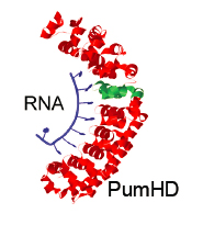
Programmable RNA-binding protein composed of repeats of a single modular unit;
Katarzyna P. Adamala*, Daniel A. Martin-Alarcon*, and Edward S. Boyden;
PNAS, 2016, 10.1073/pnas.1519368113; *equal contribution
local copy pdf
publisher website link


Biocomputing

prototyping biology

As we are approaching the limits of technological abilities of silicone microchips, biological computation is one of the alternatives for future development of computing devices.

Due to the complex and unpredictable nature of live cell biochemistry, we use simpler synthetic minimal cells to engineer and validate biological circuits. 

 

prototyping biology

TRUMPET

We engineered biocomputing platform for constructing Boolean logic gates in synthetic cell system.

TRUMPET stands for Transcriptional RNA Universal Multi-Purpose GatE PlaTform.

The platform allows constructing orthogonal universal Boolean logic gates, including layering gates to engineer complex circuits. The logic gate design tool includes sequence validation and folding tests. More at trumpet.bio


Very large-scale genetic circuit design automation

We will build liposome based synthetic minimal cell based soft-computing circuits using genetic logic gates and sequential synthetic cell fusion. The proposed research will lead to a 105 scale-up of our ability to implement information processing in cells.
Large, complex genetic circuits will be implemented in both living cells and non-living vesicles, offering extremely dense and low-power computing.  The proposed research spans the scale from computing in individual cells to that performed by multicellular ecological systems.
Here, insight is taken from software engineering to make genetics programmable. Key to our approach is refactoring genetics such that units of regulation are simple and modular and thus are able to be put together by EDA software.
In other words, it is not enough to build software to model genetics, rather it also requires building the genetics that can be put together by the software.  This has led to new design principles in modularity and insulation, which we extend here by utilizing the principles of multicellular computing.
The use of non-living cell free systems and synthetic cells is a potential path towards systems that incorporate physical materials from biology and semiconductors.

prototyping biologyOrigins of life

The earliest evolution of life included a series of transition from non-living matter, through prebiotic organic synthesis and chemical evolution, towards the Last Universal Common Ancestor of all life.
Our work focuses on the immediately-pre-life stage of evolution, when chemistry became biology.
We create synthetic minimal cells that exhibit some key properties of life, without being entirely alive. Those cells express proteins inside phospholipid liposomes, using cell-free protein expression systems. Thus, represent the latest stage of prebiotic evolution, after the establishment of the Central Dogma. Those cells do not exhibit active homeostasis, but they can maintain separate internal environment, they can grow, divide and evolve. The controllability and flexibility of those minimal cells allow us studying chemical processes underlying major transitions in evolution.
In our work, we create synthetic minimal cells expressing complex genetic pathways, with membrane proteins facilitating communication with external environment. Together, this creates a comprehensive system to study the advent of cellular processes on the boundary between prebiotic and Darwinian evolution.


Space biology

prototyping biology
Terrier Orion with synthetic cell experiment, launch at NASA Wallops, 2018 06 21

Synthetic cells can be used as a chassis for biological and biomedical experiments under the conditions of space flight: performing reliable RNA and protein production and formation of drug particles will be crucial for establishing human presence on the Moon, Mars colony and for maintaining the health of people and animals during space flights and colonization. They can be prepared in advance and stored, lyophilised, for a long time.
We have developed synthetic cell epxerimenta payload, with a team of the RockSat-C 18 program. The payload was succesfully launched in a Terrier Orion sounding rocket from NASA Wallops in June 2018, increasing the TRL of this project to 6. This was, to our knowledge, the first time cell-free protein expression experiment was performed under spaceflight conditions.

Synthetic minimal cells have been proposed as a possible tool for biomanufacturing during long-term space missions and on extraterrestrial colonies, as well as a tool for future terraforming efforts. Synthetic minimal cells are bioreactors built specifically to mimic certain aspects of cellular processes, by reconstituting specific genetic pathways, encapsulated within a biomimic membrane. Thus, synthetic minimal cell technology combines most advantages of classical in vitro studies, with added capabilities, including spatial interactions, and interactions between many scaffolded and encapsulated proteins at once.
Traditionally, the information contained in the genetic material can travel only as far and as fast as the mass of the nucleic acids being duplicated during cell division. In the case of synthetic cells, since their autonomous reproduction is unnecessary, once a winning formula for a particular synthetic cell is discovered locally, it can simply be transmitted as digital information riding on optical fibers or radio waves at high speed (and using digital compression makes high bit rates and practically zero error rates feasible with currently available apparatus). Such information can be reduced back to synthetic cells anywhere with a synthetic cell generating apparatus ‐ be it in another country, spaceship or planet.

Our work focuses on developing mission architecture: preparation, storage and experimental conditions compatible with various flight scenarios. We are building syntetic cells to model various natural processes, to study elements of biology under the conditions of spaceflight. The primary goal of this research is to gather information about how natural terrestrial organisms behave under space flight conditions, for the benefit of designing human space exploration strategies. The other goal of this research is to help refine physicochemical boundaries for biochemistry as we know it, to aid in designing life detection systems.
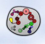

Metabolic engineering

prototyping biology

Natural cells have a whole set of endogenous metabolic processes, pathways that use small molecules for suporrting the natural cell’s metabolism and growth. One of the biggest challenges in modern metabolis engineering is to integrate newly constructed pathways with the host metabolism. When trying to discover a new pathway, or improve efficiency of an existing enzyme pathway, we often run into the problem of not being able to tell whether the changes have the desired effect in the background noise of endogenous cell metabolism.
Synthetic minimal cells have no endogenous metabolism beyond what is engineered into those cells in the first place. Therefore, we can understand and control flux of small molecule substrates within the synthetic cell. This makes designing new pathways and optimizing existing ones much easier. Additionally, the short turnaround time of a synthetic cell experiment, without needing to wait for transformants to grow to test each new plasmid combination, make iterating metabolic engineering designs much faster.
Our research focuses on engineering methods for high throughput, combinatorial design of multi-gene pathways using synthetic cells as the chassis for testing and optimizing expression.
For large scale natural product synthesis, synthetic cells are mostly research and development tool, at this point it is still cheaper and easier to perform industrial scale production in a live bug. Synthetic cell screens can help identify candidate pathways, and optimize it before scaling up for industrial manufacturing.
Synthetic cell systems can be a good tool for small scale, pilot studies, or for rapid prototyping of on-demand production of small batches of biologicals or precious small molecule compounds.


Astropharmacy

prototyping biology

During long term space missions, astronauts will encounter situations that are impossible to fully predict in advance. The isolation of the crew, for many years at a time, means that mission architecture needs to include plans for as many unpredictable situations as possible. The harsh environment of space and other planets will put high pressure on astronaut’s bodies. Providing on-demand, customizeable medical care under those conditions is very challenging. 
We cannot plan for all pharmacy needs of astronauts by stocking the required drugs in advance. Long space missions require novel approach to producing pharmacticals on short notice from pre-defined, limited raw materials.
Synthetic minimal cells provide just the right kind of programmable bioreactors that can be used to manufacture small molecule and biological drugs in short amount of time. Synthetic cells can be lyophilised and stored for a long time, and the programmability of the synthetic cell genetic circuits means almost every kind of chemical transition can be coded with the appropriate enzyme, and new enzymatic pathways for manufacturing drugs can be created on demand.
Our research focuses on designing mission-oriented solutions for preparation and storage of synthetic cells for the purposes of small scale, fast and responsive manufacturing of pharmaceuticals.


Synthetic biology

prototyping biology

Most of the efforts of biological research, both in studying healthy organisms as well as disease states, is focused either on studying the whole live cell (or organism), or on studying isolated reactions between few purified and well defined components in vitro. Most biological processes are not isolated events of interaction between few components, but rather complex interconnected networks built of often multifunctional nodes (proteins acting on many targets). The in vitro studies give results that are less relevant to natural biology – since an experiment with a few purified components does not acknowledge the vast complexity of a natural system. On the other hand, live cell studies are notoriously hard to reproduce and interpret, due to the variability between live subjects, as well as due to the inherent complexity of biology (cross-talk between the studied process and other pathways, or background signal from unrelated processes is often present).
Synthetic minimal cells deliver a solution bridging the existing gap between in vitro and live cell research: use synthetic minimal cells to investigate multicomponent gene pathways, combining the advantages of in vitro systems with the relevancy and complexity approaching that of whole cell studies.

Reading and controlling cells is the core purpose of modern synthetic biology, and the overarching goal of all biomedical studies. Both studying mechanisms of most diseases, as well as investigating healthy cellular processes, is currently done as either in vitro or live cell experiments. In vitro research methods are easy to use, cheap and efficient way to obtain information about behavior of specific, well defined protein or nucleic acid complexes or single enzymes, or to characterize small molecule interactions between metabolites or drugs and their specific biological targets. However, since life is structured in complexes that involve many components organized in precise 3D assemblies, the in vitro experiments often only deliver information about small snapshot of this complex, natural system. Studies of live cells allow to obtain truly biologically relevant information about complex pathways, but at significantly higher cost, and with results that are harder to interpret and often less reproducible. The variability between live cell subjects and the underlying intricacy of interconnected biological networks constantly interacting with each other makes signal measured in live cell experiments often more noisy and the experiment itself difficult to design.
Synthetic minimal cells offer a platform that allows studying complex genetic pathways, while keeping the complexity of the system at a level that still allows us understanding fully what the system contains and how to engineer it. Our research focuses on building tools for general use in many areas of synthetic biology, as well as studying some specific cases of complex biological processes, both healthy and diseased, that are not accessible by studying natural complex cells.


Automation

We are developing solutions for automating and scaling thropought of synthetic cell epxeriments. Microfluidic rigs help us work faster, iterating more genetic circuit designs with automated liposome formation and streamlined detection of protein expression.

Our first rig recently started producing droplets with fluorescent RNA.


Synthetic cell tools developed by our lab

We have developed a toolbox of computational resources, custom devices and protocols for engineering synthetic cells. 

Liposome calculation tools
prototyping biology


Trumpet: biocomputing platform.
prototyping biology


Liposome formation apparatus
prototyping biology


Akaby strain
prototyping biology


tRNA Foundry
prototyping biology


CyanoConstruct
prototyping biology


Addgene deposits from out lab (plasmids and bacteria strains):
www.addgene.org/Kate_Adamala/
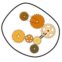


prototyping biology

Coming soon:

Cellulator


Tools are free to use; CC BY-NC-SA 4.0.
prototyping biology


Toolbox

Synthetic cell tools developed by our lab

We have developed a toolbox of computational resources, custom devices and protocols for engineering synthetic cells. 

Liposome calculation tools
prototyping biology


Trumpet: biocomputing platform.
prototyping biology


Liposome formation apparatus
prototyping biology


Akaby strain
prototyping biology


tRNA Foundry
prototyping biology


CyanoConstruct
prototyping biology


Addgene deposits from out lab (plasmids and bacteria strains):
www.addgene.org/Kate_Adamala/


prototyping biology

Coming soon:

Cellulator


Tools are free to use; CC BY-NC-SA 4.0.
prototyping biology


Liposome calculation

This spreadsheet calculates number of liposomes, number of solute molecules per liposome, % of internal volume taken by the lumen of liposomes and volume per liposome.
You can also calculate number of molecules per liposome for embedding molecules in outer leaflet of the liposome. 

To use this spreadsheet, you need to change minimum two values: lipid concentration and liposome diameter. 
Please note the units! Lipid concentration is in millimoles (mM) and liposome diameter is in nanometers (nm).  

Type the input values into the green fields.
Do not modify the blue fields unless you understand the equations and values used here. 

If you make a mistake or want to start over, refresh the page.
You can download the spreadsheet using the download icon at the bottom of the frame.

This spreadsheet is calibrated for phospholipids only. Specifically, for POPC, with surface area of 0.7nm2. We used this spreadsheet for other phospholipids too, especially diacyl lipids with modified headgroups should have similar surface area.
If you want to do calculations for fatty acids, change the surface area (nm2) value to 0.35.  

This spreadsheet makes the following assumptions: all liposomes are unilamelar, equal in size and spherical.


We would appreciate if you put our lab in acknowledgments when you use this spreadsheet for published work.


What is behind the spreadsheet

The efficiency of solute encapsulation inside POPC liposomes of a given radius r (nm) at a given concentration c (mM) can be estimated using this formula, used in the Szostak Laboratory and empirically confirmed by encapsulation experiments :

%internal volume = vol_liposome*liposomes_ml*10-19

Where:

vol_liposome = (4/3)*Pi*(r3)

is the volume of the lumen of a single liposome, in nm3;

liposomes_ml = surface_area_ml/area_liposome is the number of liposomes per 1 mL;

surface_area_ml = (c*10^-6)*((760*10^21)/0.9*NA)/2.5)/2

is the surface area of liposomes per 1 mL of solution of a given c (mM), with POPC MW=760 and length of the lipid bilayer approximated to 2.5nm; NA is Avogadro’s number;

and finally,

area_liposome = 4*Pi*(r2)

is the surface area of the liposome outer leaflet, in nm2.

These calculations were made with the assumption that liposome curvature is negligible, so the inner and outer leaflet contain an equal number of lipids and have equal surface area. The thickness of the bilayer was approximated at 2.5 nm. (1)
The addition of cholesterol increases bilayer thickness up to 30%, thus affecting the encapsulation rate (2) , but we cannot reliably estimate the influence of cholesterol on packing density and surface area of the liposomes.

According to this formula, a 25 mM solution of 200 nm POPC liposomes will contain ~14% of the total volume encapsulated inside liposomes.
In reality, the encapsulation rate of liposomes used in our experiments is likely lower. This is due to factors like the presence of cholesterol in POPC membranes and the fact, that in liposomes extruded through, e.g., a 200 nm filter, the size distribution of liposomes varies greatly and is, on average, smaller than 200nm. (3-5)
The differences in yield of protein synthesis inside synthetic cells, explained by the difference in efficiency of encapsulating the TX/TL enzyme mix, have been observed . (6)

1. Lewis, B. a & Engelman, D. M. Lipid bilayer thickness varies linearly with acyl chain length in fluid phosphatidylcholine vesicles. J. Mol. Biol. 166, 211–217 (1983).
2. Nezil, F. a. & Bloom, M. Combined influence of cholesterol and synthetic amphiphillic peptides upon bilayer thickness in model membranes. Biophys. J. 61, 1176–1183 (1992).
3. Jousma, H. et al. Characterization of liposomes. The influence of extrusion of multilamellar vesicles through polycarbonate membranes on particle size, particle size distribution and number of bilayers. Int. J. Pharm. 35, 263–274 (1987).
4. Olson, F., Hunt, C. a, Szoka, F. C., Vail, W. J. & Papahadjopoulos, D. Preparation of liposomes of defined size distribution by extrusion through polycarbonate membranes. Biochim. Biophys. Acta 557, 9–23 (1979).
5. Berger, N., Sachse, a., Bender, J., Schubert, R. & Brandl, M. Filter extrusion of liposomes using different devices: Comparison of liposome size, encapsulation efficiency, and process characteristics. Int. J. Pharm. 223, 55–68 (2001).
6. Caschera, F. & Noireaux, V. Compartmentalization of an all-E. coli Cell-Free Expression System for the Construction of a Minimal Cell. Artif. Life 22, 185-95 (2016).

learning

Learning resources

How To Grow (Almost) Anything

The HTGAA course is based on the classic Fab-Lab “Hot to Make Almost Anything” fabrication class.
BioAcademy is the best resource to learn basisc of bioengineering, including specific applications of most cutting edge techniques. We run a synthetic cell module, including overwiew of the field of synthetic life and examples of synthetic cell based tools for biological engineering.
buildacell

Build-a-Cell

prototyping biology

Build-a-Cell is an international community of scientists and policymakers working on building synthetic cells. We facilitate collaborations between groups in different areas of synthetic cell research, we engage with scientists in other disciplines to promote use of synthetic cell tools, and we provide information for the general public.
Our lab participated in every one of the bi-annual Build-a-Cell workshops, and we are actively pursuing collaborations with many Build-a-Cell members.

Kate Adamala is the co-founder and leader of Build-a-Cell. 

STAT magazine published nice overview of efforts to build artificial life, From chemicals to life: Scientists try to build cells from scratch.

Details, including list of member labs, working groups, workshops and all resources can be found at www.buildacell.org.


DIY Bio

How To Grow (Almost) Anything

The HTGAA course is based on the classic Fab-Lab “Hot to Make Almost Anything” fabrication class.
BioAcademy is the best resource to learn basisc of bioengineering, including specific applications of most cutting edge techniques. We run a synthetic cell module, including overwiew of the field of synthetic life and examples of synthetic cell based tools for biological engineering.

Biohacker 101 Class

Excellent resource for beginning and advanced DIY biological engineers, biospaces and self-taught biohackers, as well as interesting supplement for anyone studying basics of modern biological engineering.

Astrobiology

Astrobiology

Astrobiology is studying past, present and future of the life on the Universe. Synthetic cells are the perfect tool to answer many of the questions typically asked by astrobiologists, including engineering synthetic cells to resemble earliest terrestrial life forms for studying of the origins of life on Earth, engineering synthetic cell systems to explore possibilities of biochemistries yielding life on other planets, and exploring possible future evolution scenarios for various life forms. Using synthetic cells we can ask many questions about “weird life” events and alternative life forms, questions that would be nearly impossible to answer studying modern, complex terrestrial cells.

Our work is driven by the question of what is the multidimensional niche space for life, and what are the molecular mechanisms of these boundary physicochemical conditions of life? This leads naturally to the questions of how does terrestrial life responds to changes at those boundary conditions, and can synthetic biology be used to expand these? We aim at defining comprehensive matrix describing mechanisms in which environment restricts basic processes of biology, and developing framework for studying and designing living systems around those limitations.
prototyping biology

Exploring the Informational Transitions Bridging Inorganic Chemistry and Minimal Life

Understanding what separates life from non-life is critical to solving some of the great outstanding questions in science, such as how life first emerged and how we might unambiguously detect life on other worlds. The transition from non-living to living matter has been notoriously difficult to quantify, limiting our ability to develop the necessary theory for understanding life or its universal properties. One of the most significant challenges is the complexity of the examples of life available to study, which represent the product of more than 3.5 billion years of evolution. Systems representing the intermediate stages of complexity between simple chemistry and modern biology are only now becoming accessible to study in the lab with advances in systems chemistry and minimal synthetic life, respectively. These have not yet been connected to explore the uncharacterized landscape between simple chemical systems and the much more complex biochemical architectures characteristic of life today. Our project is an unprecedented opportunity to systematically study the full pathway from non-life to life through a synthesis of bottom-up chemistry, top-down minimal biology and fundamental theory. We aim to build very simple systems from chemical and biological parts, and by studying varying stages of programmed complexity and autonomy, systematically evaluate the transitions in information through which these become more life-like, self-referential entities. 

Automation

Automation

We are developing solutions for automating and scaling thropought of synthetic cell epxeriments. Microfluidic rigs help us work faster, iterating more genetic circuit designs with automated liposome formation and streamlined detection of protein expression.

Our first rig recently started producing droplets with fluorescent RNA.